
12 months from date of receipt / reconstitution, -20 °C as supplied
| 应用 | 稀释度 |
|---|---|
| WB | 1:1000 |
| ICC | 1:500 |
H3K14ac is a post-translational modification form of histone H3, specifically referring to the acetylation of its 14th lysine residue. The modification of H3K14ac is closely related to gene transcription regulation. Acetylation modifications are generally associated with the activation of gene transcription, as they can alter the chromatin structure, making DNA more accessible to transcription factors and RNA polymerases. In certain studies, such as the research on novel products of Akkermansia muciniphila, the upregulation of H3K14ac promotes the transcription and secretion of heat shock protein 70 (HSP70) in cancer cells, thereby influencing the tumor microenvironment. Abnormal modifications of H3K14ac may be related to the occurrence and development of various diseases. For instance, in epithelial ovarian tumors, the expression of H3K14Ac may be associated with the histological grade and clinical stage of ovarian cancer. In studies on arsenic poisoning, H3K14ac is linked to arsenic poisoning and arsenic-induced early toxic effects, potentially providing an effective biomarker for early monitoring of arsenic poisoning.
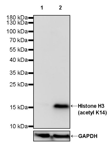
WB result of Histone H3 (acetyl K14) Recombinant Rabbit mAb
Primary antibody: Histone H3 (acetyl K14) Recombinant Rabbit mAb at 1/1000 dilution
Lane 1: untreated HeLa whole cell lysate 20 µg
Lane 2: HeLa treated with 500 ng/ml TSA for 4 hours whole cell lysate 20 µg
Secondary antibody: Goat Anti-rabbit IgG, (H+L), HRP conjugated at 1/10000 dilution
Predicted MW: 15 kDa
Observed MW: 17 kDa
WB result of Histone H3 (acetyl K14) Recombinant Rabbit mAb
Primary antibody: Histone H3 (acetyl K14) Recombinant Rabbit mAb at 1/1000 dilution
Lane 1: untreated NIH/3T3 whole cell lysate 20 µg
Lane 2: NIH/3T3 treated with 500 ng/ml TSA for 4 hours whole cell lysate 20 µg
Secondary antibody: Goat Anti-rabbit IgG, (H+L), HRP conjugated at 1/10000 dilution
Predicted MW: 15 kDa
Observed MW: 17 kDa
ICC analysis of HeLa cells treated with TSA (500ng/ml,4h) (top panel) and HeLa cells untreated with TSA (500ng/ml,4h) (below panel). Anti- Histone H3 (acetyl K14) antibody was used at 1/500 dilution (Green) and incubated overnight at 4°C. Goat polyclonal Antibody to Rabbit IgG - H&L (Alexa Fluor® 488) was used as secondary antibody at 1/1000 dilution. The cells were fixed with 100% ice-cold methanol and permeabilized with 0.1% PBS-Triton X-100. Nuclei were counterstained with DAPI (Blue). Counterstain with tubulin (Red).
 您现在的位置: