
12 months from date of receipt / reconstitution, -20 °C as supplied
| 应用 | 稀释度 |
|---|---|
| WB | 1:1000 |
| IHC-P | 1:200 |
Lysosomal-associated membrane protein 1 is a glycoprotein from a family of Lysosome-associated membrane glycoproteins. The LAMP-1 glycoprotein is a type I transmembrane protein which is expressed at high or medium levels in at least 76 different normal tissue cell types. It resides primarily across lysosomal membranes, and functions to provide selectins with carbohydrate ligands. CD107a has also been shown to be a marker of degranulation on lymphocytes such as CD8+ and NK cells, and may also play a role in tumor cell differentiation and metastasis. Although the LAMP1 glycoproteins primarily reside across lysosomal membranes, in certain cases they can be expressed across the plasma membrane of the cell. Expression of LAMP1 at the cell surface can occur due to lysosomal fusion with the cell membrane. Cell surface expression of LAMP1 can serve as a ligand for selectins and help mediate cell-cell adhesion. Accordingly, cell surface expression of LAMP1 is seen in cells with migratory or invasive functions, such as cytotoxic T cells, platelets and macrophages. Cell surface expression of LAMP1 and LAMP2 is also often seen in cancer cells, particularly cancers with high metastatic potential, such as colon carcinoma and melanoma, and has been shown to correlate with their metastatic potential.
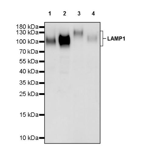
WB result of LAMP1 Recombinant Rabbit mAb
Primary antibody: LAMP1 Recombinant Rabbit mAb at 1/1000 dilution
Lane 1: HeLa whole cell lysate 20 µg
Lane 2: JAR whole cell lysate 20 µg
Lane 3: U937 whole cell lysate 20 µg
Lane 4: Jurkat whole cell lysate 20 µg
Secondary antibody: Goat Anti-rabbit IgG, (H+L), HRP conjugated at 1/10000 dilution
Predicted MW: 45 kDa
Observed MW: 90~140 kDa
This blot was developed with high sensitivity substrate
WB result of LAMP1 Recombinant Rabbit mAb
Primary antibody: LAMP1 Recombinant Rabbit mAb at 1/1000 dilution
Lane 1: mouse kidney lysate 20 µg
Secondary antibody: Goat Anti-rabbit IgG, (H+L), HRP conjugated at 1/10000 dilution
Predicted MW: 45 kDa
Observed MW: 120 kDa
IHC shows positive staining in paraffin-embedded human kidney. Anti- LAMP1 antibody was used at 1/200 dilution, followed by a HRP Polymer for Mouse & Rabbit IgG (ready to use). Counterstained with hematoxylin. Heat mediated antigen retrieval with Tris/EDTA buffer pH9.0 was performed before commencing with IHC staining protocol.
IHC shows positive staining in paraffin-embedded human ovarian cancer. Anti- LAMP1 antibody was used at 1/200 dilution, followed by a HRP Polymer for Mouse & Rabbit IgG (ready to use). Counterstained with hematoxylin. Heat mediated antigen retrieval with Tris/EDTA buffer pH9.0 was performed before commencing with IHC staining protocol.
IHC shows positive staining in paraffin-embedded human thyroid cancer. Anti- LAMP1 antibody was used at 1/200 dilution, followed by a HRP Polymer for Mouse & Rabbit IgG (ready to use). Counterstained with hematoxylin. Heat mediated antigen retrieval with Tris/EDTA buffer pH9.0 was performed before commencing with IHC staining protocol.
IHC shows positive staining in paraffin-embedded rat kidney. Anti- LAMP1 antibody was used at 1/200 dilution, followed by a HRP Polymer for Mouse & Rabbit IgG (ready to use). Counterstained with hematoxylin. Heat mediated antigen retrieval with Tris/EDTA buffer pH9.0 was performed before commencing with IHC staining protocol.
 您现在的位置: