
| 应用 | 稀释度 |
|---|---|
| FCM | 5 μl per million cells in 100μl volume |
CD80 (B7-1) is a cell surface glycoprotein that belongs to the B7 family of immune regulatory molecules. It is primarily expressed on antigen-presenting cells, such as dendritic cells, macrophages, and activated B cells. CD80 functions as a costimulatory molecule that interacts with CD28 and CTLA-4 receptors expressed on T cells. This interaction plays a crucial role in the regulation of T cell activation and immune responses. Specifically, CD80 binding to CD28 on T cells provides a costimulatory signal that enhances T cell proliferation, cytokine production, and survival. This interaction promotes the development of effective immune responses against pathogens. However, CD80 also binds to CTLA-4, which has higher affinity for CD80 compared to CD28. The CD80-CTLA-4 interaction inhibits T cell activation and proliferation, thus preventing excessive or autoreactive immune responses.
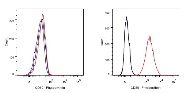
Flow cytometric analysis of Human CD80 expression on Raji. Cells from the Raji (Human Burkitt's lymphoma B lymphocyte, Right) or 293T (Human embryonic kidney epithelial cell, Left) was stained with Phycoerythrin Rabbit IgG Isotype Control (Black line histogram) and SDT PE Rabbit Anti-Human CD80 Antibody (Red line histogram) at 0.1 μg/test, cells without incubation with primary antibody and secondary antibody (Blue line histogram) was used as unlabelled control. Flow cytometry and data analysis were performed using BD FACSymphony™ A1 and FlowJo™ software.
 您现在的位置: