
12 months from date of receipt / reconstitution, -20 °C as supplied
| 应用 | 稀释度 |
|---|---|
| WB | 1:1000 |
| IHC-P | 1:250 |
| ICC | 1:500 |
MCL1 (Myeloid Cell Leukemia 1) is a protein that belongs to the Bcl-2 (B-cell lymphoma 2) family of proteins. This family of proteins plays a vital role in regulating cell death, specifically apoptosis, a process that removes unwanted or damaged cells from the body. MCL1 expression is regulated by various signaling pathways and transcription factors. Its levels can be increased in response to growth factors and cytokines, promoting cell survival. Conversely, MCL1 levels can be decreased during stress conditions or by specific targeted therapies, leading to apoptosis. MCL1 is frequently overexpressed in various types of cancers, including leukemia, lymphoma, and solid tumors. Its anti-apoptotic function allows cancer cells to survive and proliferate despite damage or stress. As a result, MCL1 has been identified as a potential target for cancer therapy. Inhibiting MCL1 activity or reducing its expression can promote apoptosis in cancer cells, leading to tumor regression.
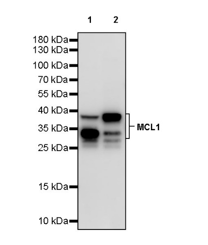
WB result of MCL1 Recombinant Rabbit mAb
Primary antibody: MCL1 Recombinant Rabbit mAb at 1/1000 dilution
Lane 1: MCF-7 whole cell lysate 20 µg
Lane 2: Raji whole cell lysate 20 µg
Secondary antibody: Goat Anti-rabbit IgG, (H+L), HRP conjugated at 1/10000 dilution
Predicted MW: 37 kDa
Observed MW: 34, 37 kDa
WB result of MCL1 Recombinant Rabbit mAb
Primary antibody: MCL1 Recombinant Rabbit mAb at 1/1000 dilution
Lane 1: rat colon lysate 20 µg
Secondary antibody: Goat Anti-rabbit IgG, (H+L), HRP conjugated at 1/10000 dilution
Predicted MW: 37 kDa
Observed MW: 30, 35 kDa
IHC shows positive staining in paraffin-embedded human tonsil. Anti-MCL1 antibody was used at 1/250 dilution, followed by a HRP Polymer for Mouse & Rabbit IgG (ready to use). Counterstained with hematoxylin. Heat mediated antigen retrieval with Tris/EDTA buffer pH9.0 was performed before commencing with IHC staining protocol.
IHC shows positive staining in paraffin-embedded human breast cancer. Anti-MCL1 antibody was used at 1/250 dilution, followed by a HRP Polymer for Mouse & Rabbit IgG (ready to use). Counterstained with hematoxylin. Heat mediated antigen retrieval with Tris/EDTA buffer pH9.0 was performed before commencing with IHC staining protocol.
IHC shows positive staining in paraffin-embedded human cervical squamous cell carcinoma. Anti-MCL1 antibody was used at 1/250 dilution, followed by a HRP Polymer for Mouse & Rabbit IgG (ready to use). Counterstained with hematoxylin. Heat mediated antigen retrieval with Tris/EDTA buffer pH9.0 was performed before commencing with IHC staining protocol.
IHC shows positive staining in paraffin-embedded human ovarian cancer. Anti-MCL1 antibody was used at 1/250 dilution, followed by a HRP Polymer for Mouse & Rabbit IgG (ready to use). Counterstained with hematoxylin. Heat mediated antigen retrieval with Tris/EDTA buffer pH9.0 was performed before commencing with IHC staining protocol.
IHC shows positive staining in paraffin-embedded rat colon. Anti-MCL1 antibody was used at 1/250 dilution, followed by a HRP Polymer for Mouse & Rabbit IgG (ready to use). Counterstained with hematoxylin. Heat mediated antigen retrieval with Tris/EDTA buffer pH9.0 was performed before commencing with IHC staining protocol.
ICC shows high expression in MCF-7 cells (top panel) and low expression in SK-OV-3 cells (below panel). Anti-MCL1 antibody was used at 1/500 dilution (Green) and incubated overnight at 4°C. Goat polyclonal Antibody to Rabbit IgG - H&L (Alexa Fluor® 488) was used as secondary antibody at 1/1000 dilution. The cells were fixed with 100% ice-cold methanol and permeabilized with 0.1% PBS-Triton X-100. Nuclei were counterstained with DAPI (Blue). Counterstain with tubulin (Red).
 您现在的位置: