
PBS, 40% Glycerol, 0.05% BSA, 0.03% Proclin 300
12 months from date of receipt / reconstitution, -20 °C as supplied
| 应用 | 稀释度 |
|---|---|
| Dot Blot | 1:1000 |
| WB | 1:1000 |
| ICC | 1:100 |
Stat1 (Signal Transducer and Activator of Transcription 1) is a crucial signaling molecule that plays a pivotal role in cells. Upon stimulation by external factors such as cytokines and growth factors, Stat1 is activated through tyrosine kinases (JAK) and mitogen-activated protein kinases (MAPK), leading to the phosphorylation of tyrosine and serine residues at its C-terminus. Phosphorylated Stat1 forms dimers and translocates into the nucleus, binding to specific DNA sequences to activate the transcription of target genes. Stat1 plays a crucial role in the immune system as a key component of the interferon signaling pathway. It regulates cell proliferation, apoptosis, and differentiation, affecting the growth and migration of tumor cells. Phospho-Stat1 (Tyr701) is a phosphorylated form of Stat1. It results from the phosphorylation of Stat1 at the Tyr701 residue. This phosphorylation event is typically induced by cytokines and growth factors through JAK (Janus kinase) or MAPK (Mitogen-Activated Protein Kinase) signaling pathways. In the nucleus, Phospho-Stat1 (Tyr701) dimers bind to specific DNA sequences known as gamma-activated sequence (GAS) elements in the promoters of target genes. Binding to these sequences leads to the transcriptional activation of those genes, thereby influencing cellular responses to cytokines and growth factors.
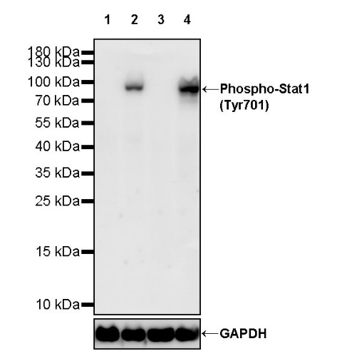
WB result of Phospho-Stat1 (Tyr701) Recombinant Rabbit mAb
Primary antibody: Phospho-Stat1 (Tyr701) Recombinant Rabbit mAb at 1/1000 dilution
Lane 1: untreated HeLa whole cell lysate 20 µg
Lane 2: HeLa treated with 100 ng/ml IFN-γ for 30 minutes whole cell lysate 20 µg
Lane 3: untreated Jurkat whole cell lysate 20 µg
Lane 4: Jurkat treated with 50 ng/ml IFN-α for 30 minutes whole cell lysate 20 µg
Secondary antibody: Goat Anti-rabbit IgG, (H+L), HRP conjugated at 1/10000 dilution
Predicted MW: 87 kDa
Observed MW: 90 kDa
WB result of Phospho-Stat1 (Tyr701) Recombinant Rabbit mAb
Primary antibody: Phospho-Stat1 (Tyr701) Recombinant Rabbit mAb at 1/1000 dilution
Lane 1: untreated PC-12 whole cell lysate 20 µg
Lane 2: PC-12 treated with 10 ng/ml hIFN-α for 30 minutes whole cell lysate 20 µg
Secondary antibody: Goat Anti-rabbit IgG, (H+L), HRP conjugated at 1/10000 dilution
Predicted MW: 87 kDa
Observed MW: 90 kDa
Dot blot result of Phospho-Stat1 (Tyr701) Recombinant Rabbit mAb
Lane1: Stat1 (Tyr701) phospho peptide
Lane2: Stat1 non-phospho peptide
Primary antibody: Phospho-Stat1 (Tyr701) Recombinant Rabbit mAb at 1/1000 dilution
Secondary antibody: Goat Anti-Rabbit IgG, (H+L), HRP conjugated at 1/10000 dilution
ICC analysis of HeLa cells treated with IFN-γ (100ng/mL, 30min) (top panel) and HeLa cells untreated with IFN-γ (100ng/mL, 30min) (below panel). Anti- Phospho-Stat1 (Tyr701) antibody was used at 1/100 dilution (Green) and incubated overnight at 4°C. Goat polyclonal Antibody to Rabbit IgG - H&L (Alexa Fluor® 488) was used as secondary antibody at 1/1000 dilution. The cells were fixed with 100% ice-cold methanol and permeabilized with 0.1% PBS-Triton X-100. Nuclei were counterstained with DAPI (Blue). Counterstain with tubulin (Red).
 您现在的位置: