
12 months from date of receipt / reconstitution, -20 °C as supplied
| 应用 | 稀释度 |
|---|---|
| WB | 1:1000 |
| ICC | 1:500 |
| ICFCM | 1:50 |
YAP1, also known as YAP (Yes-Associated Protein) or WWTR1 (WW Domain Containing Transcription Regulator 1), is a transcriptional co-activator that plays a pivotal role in the Hippo signaling pathway. When YAP1 is in an active state (commonly referred to as active YAP1), it enters the nucleus and binds to transcription factors called TEADs (TEA Domain Family Members). This YAP-TEAD complex then promotes the expression of a series of genes associated with cell proliferation, survival, and migration. However, when the Hippo signaling pathway is activated, the activity of YAP1 is suppressed. This often involves the phosphorylation of YAP1, which leads to its binding with 14-3-3 proteins and retention in the cytoplasm, preventing it from entering the nucleus to activate transcription. Abnormal expression or dysfunction of active YAP1 has been linked to various diseases, including cancer, heart disease, and neurological disorders. Therefore, studying the regulatory mechanisms of YAP1 activity and its role in diseases is crucial for developing new therapeutic strategies.
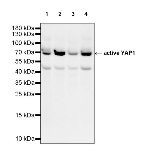
WB result of active YAP1 Recombinant Rabbit mAb
Primary antibody: active YAP1 Recombinant Rabbit mAb at 1/1000 dilution
Lane 1: HeLa whole cell lysate 20 µg
Lane 2: MCF7 whole cell lysate 20 µg
Lane 3: A549 whole cell lysate 20 µg
Lane 4: HaCaT whole cell lysate 20 µg
Secondary antibody: Goat Anti-rabbit IgG, (H+L), HRP conjugated at 1/10000 dilution
Predicted MW: 65~78 kDa
Observed MW: 65~78 kDa
Flow cytometric analysis of 4% PFA fixed 90% methanol permeabilized A549 (Human lung carcinoma epithelial cell) labelling active YAP1 antibody at 1/50 dilution (1 μg)/ (Red) compared with a Rabbit monoclonal IgG (Black) isotype control and an unlabelled control (cells without incubation with primary antibody and secondary antibody) (Blue). Goat Anti - Rabbit IgG Alexa Fluor® 488 was used as the secondary antibody.
ICC shows positive staining in A549 cells. Anti- active YAP1 antibody was used at 1/500 dilution (Green) and incubated overnight at 4°C. Goat polyclonal Antibody to Rabbit IgG - H&L (Alexa Fluor® 488) was used as secondary antibody at 1/1000 dilution. The cells were fixed with 4% PFA and permeabilized with 0.1% PBS-Triton X-100. Nuclei were counterstained with DAPI (Blue). Counterstain with tubulin (Red).
 您现在的位置: