
12 months from date of receipt / reconstitution, -20 °C as supplied
| 应用 | 稀释度 |
|---|---|
| WB | 1:1000 |
| IHC-P | 1:200 |
| ICC | 1:500 |
| FCM | 1:500 |
The CD44 antigen is a cell-surface glycoprotein involved in cell–cell interactions, cell adhesion and migration. CD44 is expressed in a large number of mammalian cell types. The standard isoform, designated CD44s, comprising exons 1–5 and 16–20 is expressed in most cell types. CD44 splice variants containing variable exons are designated CD44v. Some epithelial cells also express a larger isoform (CD44E), which includes exons v8–10. CD44 is a receptor for hyaluronic acid and internalizes metals bound to hyaluronic acid and can also interact with other ligands, such as osteopontin, collagens, and matrix metalloproteinases (MMPs). CD44, along with CD25, is used to track early T cell development in the thymus. D44 expression is an indicative marker for effector-memory T-cells. Memory cell proliferation (activation) can also be assayed in vitro with CFSE chemical tagging. In addition, variations in CD44 are reported as cell surface markers for some breast and prostate cancer stem cells. In breast cancer research CD44+/CD24- expression is commonly used as a marker for breast CSCs and is used to sort breast cancer cells into a population enriched in cells with stem-like characteristics and has been seen as an indicator of increased survival time in epithelial ovarian cancer patients.
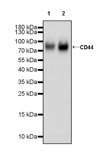
WB result of CD44 Recombinant Rabbit mAb
Primary antibody: CD44 Recombinant Rabbit mAb at 1/1000 dilution
Lane 1: A549 whole cell lysate 20 µg
Lane 2: HeLa whole cell lysate 20 µg
Secondary antibody: Goat Anti-rabbit IgG, (H+L), HRP conjugated at 1/10000 dilution
Predicted MW: 81 kDa
Observed MW: 85 kDa
Flow cytometric analysis of Daudi (Human Burkitt's lymphoma lymphoblast, left) / A431 (Human epidermoid carcinoma epithelial cell, Right) labelling CD44s antibody at 1/500 dilution (0.1 μg) / (Red) compared with a Rabbit monoclonal IgG (Black) isotype control and an unlabelled control (cells without incubation with primary antibody and secondary antibody) (Blue). Goat Anti - Rabbit IgG Alexa Fluor® 488 was used as the secondary antibody.
Negative control: Daudi
IHC shows positive staining in paraffin-embedded human tonsil. Anti-CD44 antibody was used at 1/200 dilution, followed by a HRP Polymer for Mouse & Rabbit IgG (ready to use). Counterstained with hematoxylin. Heat mediated antigen retrieval with Tris/EDTA buffer pH9.0 was performed before commencing with IHC staining protocol.
IHC shows positive staining in paraffin-embedded human spleen. Anti-CD44 antibody was used at 1/200 dilution, followed by a HRP Polymer for Mouse & Rabbit IgG (ready to use). Counterstained with hematoxylin. Heat mediated antigen retrieval with Tris/EDTA buffer pH9.0 was performed before commencing with IHC staining protocol.
IHC shows positive staining in paraffin-embedded human prostate. Anti-CD44 antibody was used at 1/200 dilution, followed by a HRP Polymer for Mouse & Rabbit IgG (ready to use). Counterstained with hematoxylin. Heat mediated antigen retrieval with Tris/EDTA buffer pH9.0 was performed before commencing with IHC staining protocol.
IHC shows positive staining in paraffin-embedded human transitional cell carcinoma. Anti-CD44 antibody was used at 1/200 dilution, followed by a HRP Polymer for Mouse & Rabbit IgG (ready to use). Counterstained with hematoxylin. Heat mediated antigen retrieval with Tris/EDTA buffer pH9.0 was performed before commencing with IHC staining protocol.
IHC shows positive staining in paraffin-embedded human breast cancer. Anti-CD44 antibody was used at 1/200 dilution, followed by a HRP Polymer for Mouse & Rabbit IgG (ready to use). Counterstained with hematoxylin. Heat mediated antigen retrieval with Tris/EDTA buffer pH9.0 was performed before commencing with IHC staining protocol.
IHC shows positive staining in paraffin-embedded human cervical squamous cell carcinoma. Anti-CD44 antibody was used at 1/200 dilution, followed by a HRP Polymer for Mouse & Rabbit IgG (ready to use). Counterstained with hematoxylin. Heat mediated antigen retrieval with Tris/EDTA buffer pH9.0 was performed before commencing with IHC staining protocol.
IHC shows positive staining in paraffin-embedded human pancreatic carcinoma. Anti-CD44 antibody was used at 1/200 dilution, followed by a HRP Polymer for Mouse & Rabbit IgG (ready to use). Counterstained with hematoxylin. Heat mediated antigen retrieval with Tris/EDTA buffer pH9.0 was performed before commencing with IHC staining protocol.
ICC shows positive staining in A431 cells (top panel) and negative staining in Daudi cells (below panel). Anti-CD44 antibody was used at 1/500 dilution (Green) and incubated overnight at 4°C. Goat polyclonal Antibody to Rabbit IgG - H&L (Alexa Fluor® 488) was used as secondary antibody at 1/1000 dilution. The cells were fixed with 100% ice-cold methanol and permeabilized with 0.1% PBS-Triton X-100. Nuclei were counterstained with DAPI (Blue). Counterstain with tubulin (Red).
 您现在的位置: