
12 months from date of receipt / reconstitution, -20 °C as supplied
| 应用 | 稀释度 |
|---|---|
| WB | 1:1000 |
| IP | 1:50 |
| IHC-P | 1:250 |
Neuropeptide Y (NPY) is a 36 amino-acid neuropeptide that is involved in various physiological and homeostatic processes in both the central and peripheral nervous systems. It is secreted alongside other neurotransmitters such as GABA and glutamate. Neuropeptide Y has been identified as being synthesized in GABAergic neurons and to act as a neurotransmitter during cellular communication. Neuropeptide Y is expressed in interneurons. NPY exerts most of its effects through Neuropeptide Y receptors, mainly Y1, Y2, Y4, and Y6. High concentrations of neuropeptide Y synthesis and action have been found in the hypothalamus and hippocampus, specifically in the arcuate nucleus (ARC) and dentate gyrus. The arcuate nucleus has been found to have one of the highest concentrations of NPY. This allows NPY to regulate neuroendocrine release of various hypothalamic hormones such as luteinizing hormone. NPY is able to modulate the mitochondrial network by affecting the expression of many genes involved in mitochondrial functions and dynamics. Neuropeptide Y has been indicated as playing an important role in neurogenesis in various parts of the brain. Two particular brain areas where NPY affects neurogenesis are the sub-ventricular zone and the dentate gyrus of the hippocampus. These areas are where cell growth and proliferation occur into adulthood.
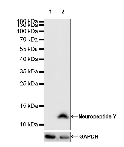
WB result of Neuropeptide Y Recombinant Rabbit mAb
Primary antibody: Neuropeptide Y Recombinant Rabbit mAb at 1/1000 dilution
Lane 1: untreated SH-SY5Y whole cell lysate 20 µg
Lane 2: SH-SY5Y treated with 200 nM TPA for 24 hours whole cell lysate 20 µg
Secondary antibody: Goat Anti-rabbit IgG, (H+L), HRP conjugated at 1/10000 dilution
Predicted MW: 11 kDa
Observed MW: 11 kDa
This blot was developed with high sensitivity substrate
WB result of Neuropeptide Y Recombinant Rabbit mAb
Primary antibody: Neuropeptide Y Recombinant Rabbit mAb at 1/1000 dilution
Lane 1: rat spleen lysate 20 µg
Secondary antibody: Goat Anti-rabbit IgG, (H+L), HRP conjugated at 1/10000 dilution
Predicted MW: 11 kDa
Observed MW: 11 kDa
This blot was developed with high sensitivity substrate
Neuropeptide Y Rabbit mAb at 1/50 dilution (1 µg) immunoprecipitating Neuropeptide Y in 0.4 mg SH-SY5Y treated with 200 nM TPA for 24 hours whole cell lysate.
Western blot was performed on the immunoprecipitate using Neuropeptide Y Rabbit mAb at 1/1000 dilution.
Secondary antibody (HRP) for IP was used at 1/1000 dilution.
Lane 1: SH-SY5Y treated with 200 nM TPA for 24 hours whole cell lysate 20 µg (Input)
Lane 2: Neuropeptide Y Rabbit mAb IP in SH-SY5Y treated with 200 nM TPA for 24 hours whole cell lysate
Lane 3: Rabbit monoclonal IgG IP in SH-SY5Y treated with 200 nM TPA for 24 hours whole cell lysate
Predicted MW: 11 kDa
Observed MW: 11 kDa
This blot was developed with high sensitivity substrate
IHC shows positive staining in paraffin-embedded human cerebral cortex. Anti- Neuropeptide Y antibody was used at 1/250 dilution, followed by a HRP Polymer for Mouse & Rabbit IgG (ready to use). Counterstained with hematoxylin. Heat mediated antigen retrieval with Tris/EDTA buffer pH9.0 was performed before commencing with IHC staining protocol.
IHC shows positive staining in paraffin-embedded human prostatic cancer. Anti- Neuropeptide Y antibody was used at 1/250 dilution, followed by a HRP Polymer for Mouse & Rabbit IgG (ready to use). Counterstained with hematoxylin. Heat mediated antigen retrieval with Tris/EDTA buffer pH9.0 was performed before commencing with IHC staining protocol.
Negative control: IHC shows negative staining in paraffin-embedded human skeletal muscle. Anti- Neuropeptide Y antibody was used at 1/250 dilution, followed by a HRP Polymer for Mouse & Rabbit IgG (ready to use). Counterstained with hematoxylin. Heat mediated antigen retrieval with Tris/EDTA buffer pH9.0 was performed before commencing with IHC staining protocol.
IHC shows positive staining in paraffin-embedded mouse cerebral cortex. Anti- Neuropeptide Y antibody was used at 1/250 dilution, followed by a HRP Polymer for Mouse & Rabbit IgG (ready to use). Counterstained with hematoxylin. Heat mediated antigen retrieval with Tris/EDTA buffer pH9.0 was performed before commencing with IHC staining protocol.
IHC shows positive staining in paraffin-embedded rat cerebral cortex. Anti- Neuropeptide Y antibody was used at 1/250 dilution, followed by a HRP Polymer for Mouse & Rabbit IgG (ready to use). Counterstained with hematoxylin. Heat mediated antigen retrieval with Tris/EDTA buffer pH9.0 was performed before commencing with IHC staining protocol.
 您现在的位置: