
12 months from date of receipt / reconstitution, -20 °C as supplied
| 应用 | 稀释度 |
|---|---|
| WB | 1:1000 |
| IP | 1:50 |
| IHC-P | 1:1000 |
| IF | 1:1000 |
The RPE65 protein, also known as Retinoid isomerohydrolase, is a crucial protein produced in the retinal pigment epithelium (RPE) cells. PE65 protein serves as a key isomerohydrolase in the visual cycle, involved in the regeneration of 11-cis-retinal, which is the chromophore of rod and cone cell visual pigments. It catalyzes the cleavage and isomerization of all-trans-retinyl fatty acid esters to produce 11-cis-retinol, which is further oxidized by 11-cis-retinol dehydrogenase to 11-cis-retinal. RPE65 protein exists in two forms: soluble sRPE65 and membrane-bound mRPE65. The latter is palmitoylated and serves as a palmitoyl donor for lecithin retinol acyltransferase (LRAT), which catalyzes a step in the regeneration of vitamin A to all-trans-retinol.
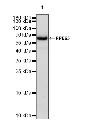
WB result of RPE65 Recombinant Rabbit mAb
Primary antibody: RPE65 Recombinant Rabbit mAb at 1/1000 dilution
Lane 1: mouse eye lysate 20 µg
Secondary antibody: Goat Anti-rabbit IgG, (H+L), HRP conjugated at 1/10000 dilution
Predicted MW: 61 kDa
Observed MW: 65 kDa
WB result of RPE65 Recombinant Rabbit mAb
Primary antibody: RPE65 Recombinant Rabbit mAb at 1/1000 dilution
Lane 1: rat eye lysate 20 µg
Secondary antibody: Goat Anti-rabbit IgG, (H+L), HRP conjugated at 1/10000 dilution
Predicted MW: 61 kDa
Observed MW: 65 kDa
RPE65 Rabbit mAb at 1/50 dilution (1 µg) immunoprecipitating RPE65 in 0.4 mg mouse eye lysate.
Western blot was performed on the immunoprecipitate using RPE65 Rabbit mAb at 1/1000 dilution.
Secondary antibody (HRP) for IP was used at 1/1000 dilution.
Lane 1: mouse eye lysate 20 µg (Input)
Lane 2: RPE65 Rabbit mAb IP in mouse eye lysate
Lane 3: Rabbit monoclonal IgG IP in mouse eye lysate
Predicted MW: 61 kDa
Observed MW: 65 kDa
IHC shows positive staining in paraffin-embedded mouse retina. Anti-RPE65 antibody was used at 1/1000 dilution, followed by a HRP Polymer for Mouse & Rabbit IgG (ready to use). Counterstained with hematoxylin. Heat mediated antigen retrieval with Tris/EDTA buffer pH9.0 was performed before commencing with IHC staining protocol.
IHC shows positive staining in paraffin-embedded rat retina. Anti-RPE65 antibody was used at 1/1000 dilution, followed by a HRP Polymer for Mouse & Rabbit IgG (ready to use). Counterstained with hematoxylin. Heat mediated antigen retrieval with Tris/EDTA buffer pH9.0 was performed before commencing with IHC staining protocol.
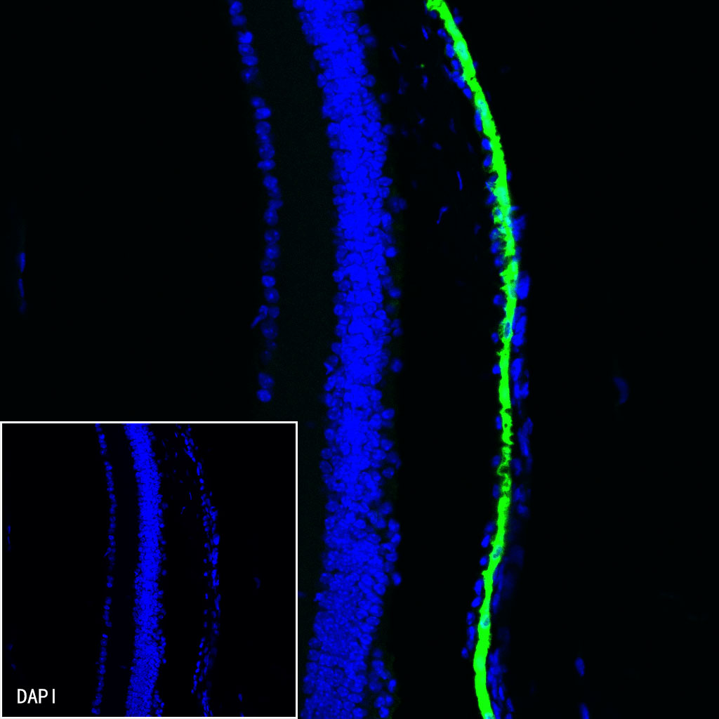
IF shows positive staining in paraffin-embedded mouse retina. Anti-RPE65 antibody was used at 1/1000 dilution (Green) and incubated overnight at 4°C. Goat polyclonal Antibody to Rabbit IgG - H&L (Alexa Fluor® 488) was used as secondary antibody at 1/1000 dilution. Counterstained with DAPI (Blue). Heat mediated antigen retrieval with EDTA buffer pH9.0 was performed before commencing with IF staining protocol.

IF shows positive staining in paraffin-embedded rat retina. Anti-RPE65 antibody was used at 1/1000 dilution (Green) and incubated overnight at 4°C. Goat polyclonal Antibody to Rabbit IgG - H&L (Alexa Fluor® 488) was used as secondary antibody at 1/1000 dilution. Counterstained with DAPI (Blue). Heat mediated antigen retrieval with EDTA buffer pH9.0 was performed before commencing with IF staining protocol.
 您现在的位置: