
PBS, 40% Glycerol, 0.05% BSA, 0.03% Proclin 300
12 months from date of receipt / reconstitution, -20 °C as supplied
| 应用 | 稀释度 |
|---|---|
| WB | 1:1000 |
| IHC-P | 1:500 |
| ICC | 1:500 |
| ICFCM | 1:500 |
| ChIP | 1:20-1:50 |
Histone H3 (mono-methylated K36) refers to a specific form of histone H3 where the lysine residue at position 36 (K36) undergoes a monomethylation modification. This epigenetic modification, known as histone methylation, can influence the interaction between histones and DNA, thereby regulating gene transcription activity without altering the DNA sequence. The mono-methylation of H3K36 can be associated with either gene activation or repression, depending on the context and extent of methylation. In biological research, Histone H3 (mono-methylated K36) is often used as a tool or marker to study epigenetics, transcriptional regulation, cell differentiation, and other biological processes. Additionally, in disease research, abnormal patterns of histone methylation, including H3K36 methylation, may be associated with the development and progression of certain diseases.
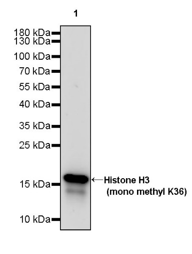
WB result of Histone H3 (mono methyl K36) Recombinant Rabbit mAb
Primary antibody: Histone H3 (mono methyl K36) Rabbit mAb at 1/1000 dilution
Lane 1: HeLa whole cell lysate 20 µg
Secondary antibody: Goat Anti-rabbit IgG, (H+L), HRP conjugated at 1/10000 dilution
Predicted MW: 15 kDa
Observed MW: 17 kDa
WB result of Histone H3 (mono methyl K36) Recombinant Rabbit mAb
Primary antibody: Histone H3 (mono methyl K36) Rabbit mAb at 1/1000 dilution
Lane 1: NIH/3T3 whole cell lysate 20 µg
Secondary antibody: Goat Anti-rabbit IgG, (H+L), HRP conjugated at 1/10000 dilution
Predicted MW: 15 kDa
Observed MW: 17 kDa
Flow cytometric analysis of 4% PFA fixed 90% methanol permeabilized HeLa (Human cervix adenocarcinoma epithelial cell) labelling Histone H3 (mono methyl K36) antibody at 1/500 dilution (0.1 μg)/ (Red) compared with a Rabbit monoclonal IgG (Black) isotype control and an unlabelled control (cells without incubation with primary antibody and secondary antibody) (Blue). Goat Anti - Rabbit IgG Alexa Fluor® 488 was used as the secondary antibody.
Flow cytometric analysis of 4% PFA fixed 90% methanol permeabilized NIH/3T3 (Mouse embryonic fibroblast) labelling Histone H3 (mono methyl K36) antibody at 1/500 dilution (0.1 μg)/ (Red) compared with a Rabbit monoclonal IgG (Black) isotype control and an unlabelled control (cells without incubation with primary antibody and secondary antibody) (Blue). Goat Anti - Rabbit IgG Alexa Fluor® 488 was used as the secondary antibody.
IHC shows positive staining in paraffin-embedded human stomach. Anti-Histone H3 (mono methyl K36) antibody was used at 1/500 dilution, followed by a HRP Polymer for Mouse & Rabbit IgG (ready to use). Counterstained with hematoxylin. Heat mediated antigen retrieval with Tris/EDTA buffer pH9.0 was performed before commencing with IHC staining protocol.
IHC shows positive staining in paraffin-embedded human testis. Anti-Histone H3 (mono methyl K36) antibody was used at 1/500 dilution, followed by a HRP Polymer for Mouse & Rabbit IgG (ready to use). Counterstained with hematoxylin. Heat mediated antigen retrieval with Tris/EDTA buffer pH9.0 was performed before commencing with IHC staining protocol.
IHC shows positive staining in paraffin-embedded mouse colon. Anti-Histone H3 (mono methyl K36) antibody was used at 1/500 dilution, followed by a HRP Polymer for Mouse & Rabbit IgG (ready to use). Counterstained with hematoxylin. Heat mediated antigen retrieval with Tris/EDTA buffer pH9.0 was performed before commencing with IHC staining protocol.
IHC shows positive staining in paraffin-embedded mouse lung. Anti-Histone H3 (mono methyl K36) antibody was used at 1/500 dilution, followed by a HRP Polymer for Mouse & Rabbit IgG (ready to use). Counterstained with hematoxylin. Heat mediated antigen retrieval with Tris/EDTA buffer pH9.0 was performed before commencing with IHC staining protocol.
IHC shows positive staining in paraffin-embedded rat colon. Anti-Histone H3 (mono methyl K36) antibody was used at 1/500 dilution, followed by a HRP Polymer for Mouse & Rabbit IgG (ready to use). Counterstained with hematoxylin. Heat mediated antigen retrieval with Tris/EDTA buffer pH9.0 was performed before commencing with IHC staining protocol.
IHC shows positive staining in paraffin-embedded rat liver. Anti-Histone H3 (mono methyl K36) antibody was used at 1/500 dilution, followed by a HRP Polymer for Mouse & Rabbit IgG (ready to use). Counterstained with hematoxylin. Heat mediated antigen retrieval with Tris/EDTA buffer pH9.0 was performed before commencing with IHC staining protocol.
ICC shows positive staining in HeLa cells. Anti- Histone H3 (mono methyl K36) antibody was used at 1/500 dilution (Green) and incubated overnight at 4°C. Goat polyclonal Antibody to Rabbit IgG - H&L (Alexa Fluor® 488) was used as secondary antibody at 1/1000 dilution. The cells were fixed with 100% ice-cold methanol and permeabilized with 0.1% PBS-Triton X-100. Nuclei were counterstained with DAPI (Blue). Counterstain with tubulin (Red).
ICC shows positive staining in NIH/3T3 cells. Anti- Histone H3 (mono methyl K36) antibody was used at 1/500 dilution (Green) and incubated overnight at 4°C. Goat polyclonal Antibody to Rabbit IgG - H&L (Alexa Fluor® 488) was used as secondary antibody at 1/1000 dilution. The cells were fixed with 4% PFA and permeabilized with 0.1% PBS-Triton X-100. Nuclei were counterstained with DAPI (Blue). Counterstain with tubulin (Red).
Chromatin immunoprecipitation (ChIP) was performed on HeLa cells cross - linked with 1% formaldehyde for 10 min, then chromatin was fragmented by sonication. Parallel reactions used Histone H3 (mono methyl K36) Recombinant Rabbit mAb (S-R396) and Rabbit mAb IgG Isotype Control (SDT-R173) at 1:50 for immunoprecipitation.
Post - immunoprecipitation, both samples were washed, eluted, and cross - links reversed. Purified DNA was analyzed by qPCR.
qPCR (%input: immunoprecipitated DNA/input DNA) showed the enrichment of RPL30, GAPDH, MYOD1, AMF, SAT-α and SAT-2 in Histone H3 (mono methyl K36) Recombinant Rabbit mAb (S-R396) - immunoprecipitated sample.
 您现在的位置: