
12 months from date of receipt / reconstitution, -20 °C as supplied
| 应用 | 稀释度 |
|---|---|
| WB | 1:1000 |
| IHC-P | 1:500-1:2000 |
HLA-E is a non-classical MHC class I molecule that exhibits limited polymorphism and lower cell surface expression compared to its classical counterparts. HLA-E plays a unique role in immune responses. It can present antigen epitopes derived from pathogens, tumors, and self-antigens to CD8+ T cells for recognition by their T cell receptors (TCRs), mediating CD8+ T cell responses. HLA-E can interact with inhibitory receptors CD94/NKG2A/C on NK cells and some T cells, regulating their killing activities. Recent research has found that HLA-E-restricted T cells are able to recognize and destroy infected cells, playing a crucial role in the immune response against COVID-19. HLA-E was found to be resistant to inhibition by the virus, even though other classical HLA proteins were suppressed. When HLA-E-restricted T cells recognize the presented antigen fragments, they become activated and release cytotoxic factors to destroy the infected cells.
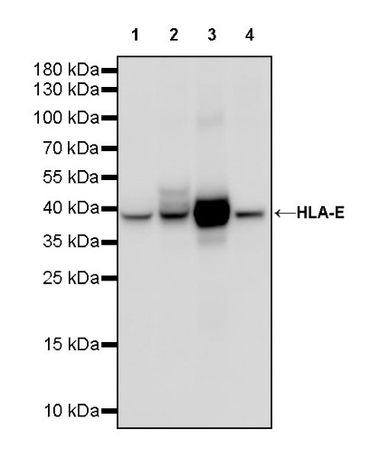
WB result of HLA-E Rabbit mAb
Primary antibody: HLA-E Rabbit mAb at 1/1000 dilution
Lane 1: THP-1 whole cell lysate 20 µg
Lane 2: Jurkat whole cell lysate 20 µg
Lane 3: HL-60 whole cell lysate 20 µg
Lane 4: Daudi whole cell lysate 20 µg
Secondary antibody: Goat Anti-rabbit IgG, (H+L), HRP conjugated at 1/10000 dilution
Predicted MW: 40 kDa
Observed MW: 40 kDa
WB result of HLA-E Rabbit mAb
Primary antibody: HLA-E Rabbit mAb at 1/1000 dilution
Lane 1: mouse spleen lysate 20 µg
Secondary antibody: Goat Anti-rabbit IgG, (H+L), HRP conjugated at 1/10000 dilution
Predicted MW: 40 kDa
Observed MW: 50 kDa
IHC shows positive staining in paraffin-embedded human liver. Anti- HLA-E antibody was used at 1/2000 dilution, followed by a HRP Polymer for Mouse & Rabbit IgG (ready to use). Counterstained with hematoxylin. Heat mediated antigen retrieval with Tris/EDTA buffer pH9.0 was performed before commencing with IHC staining protocol.
IHC shows positive staining in paraffin-embedded human cerebral cortex. Anti- HLA-E antibody was used at 1/2000 dilution, followed by a HRP Polymer for Mouse & Rabbit IgG (ready to use). Counterstained with hematoxylin. Heat mediated antigen retrieval with Tris/EDTA buffer pH9.0 was performed before commencing with IHC staining protocol.
IHC shows positive staining in paraffin-embedded human kidney. Anti- HLA-E antibody was used at 1/2000 dilution, followed by a HRP Polymer for Mouse & Rabbit IgG (ready to use). Counterstained with hematoxylin. Heat mediated antigen retrieval with Tris/EDTA buffer pH9.0 was performed before commencing with IHC staining protocol.
IHC shows positive staining in paraffin-embedded human spleen. Anti- HLA-E antibody was used at 1/2000 dilution, followed by a HRP Polymer for Mouse & Rabbit IgG (ready to use). Counterstained with hematoxylin. Heat mediated antigen retrieval with Tris/EDTA buffer pH9.0 was performed before commencing with IHC staining protocol.
IHC shows positive staining in paraffin-embedded human tonsil. Anti- HLA-E antibody was used at 1/2000 dilution, followed by a HRP Polymer for Mouse & Rabbit IgG (ready to use). Counterstained with hematoxylin. Heat mediated antigen retrieval with Tris/EDTA buffer pH9.0 was performed before commencing with IHC staining protocol.
IHC shows positive staining in paraffin-embedded human ovarian carcinoma Anti- HLA-E antibody was used at 1/500 dilution, followed by a HRP Polymer for Mouse & Rabbit IgG (ready to use). Counterstained with hematoxylin. Heat mediated antigen retrieval with Tris/EDTA buffer pH9.0 was performed before commencing with IHC staining protocol.
IHC shows positive staining in paraffin-embedded human gastric cancer. Anti- HLA-E antibody was used at 1/500 dilution, followed by a HRP Polymer for Mouse & Rabbit IgG (ready to use). Counterstained with hematoxylin. Heat mediated antigen retrieval with Tris/EDTA buffer pH9.0 was performed before commencing with IHC staining protocol.
IHC shows positive staining in paraffin-embedded human transitional cell carcinoma Anti- HLA-E antibody was used at 1/500 dilution, followed by a HRP Polymer for Mouse & Rabbit IgG (ready to use). Counterstained with hematoxylin. Heat mediated antigen retrieval with Tris/EDTA buffer pH9.0 was performed before commencing with IHC staining protocol.
IHC shows positive staining in paraffin-embedded mouse colon. Anti- HLA-E antibody was used at 1/500 dilution, followed by a HRP Polymer for Mouse & Rabbit IgG (ready to use). Counterstained with hematoxylin. Heat mediated antigen retrieval with Tris/EDTA buffer pH9.0 was performed before commencing with IHC staining protocol.
IHC shows positive staining in paraffin-embedded mouse liver. Anti- HLA-E antibody was used at 1/500 dilution, followed by a HRP Polymer for Mouse & Rabbit IgG (ready to use). Counterstained with hematoxylin. Heat mediated antigen retrieval with Tris/EDTA buffer pH9.0 was performed before commencing with IHC staining protocol.
 您现在的位置: