
12 months from date of receipt / reconstitution, -20 °C as supplied
| 应用 | 稀释度 |
|---|---|
| WB | 1:1000 |
| IP | 1:50 |
| ICC | 1:500 |
Interleukin 8 (IL-8 or chemokine (C-X-C motif) ligand 8, CXCL8) is a chemokine produced by macrophages and other cell types such as epithelial cells, airway smooth muscle cells and endothelial cells. Endothelial cells store IL-8 in their storage vesicles, the Weibel-Palade bodies. IL-8 has two primary functions. It induces chemotaxis in target cells, primarily neutrophils but also other granulocytes, causing them to migrate toward the site of infection. IL-8 also stimulates phagocytosis once they have arrived. IL-8 is also known to be a potent promoter of angiogenesis. In target cells, IL-8 induces a series of physiological responses required for migration and phagocytosis, such as increases in intracellular Ca2+, exocytosis (e.g. histamine release), and the respiratory burst. Interleukin-8 is a key mediator associated with inflammation where it plays a key role in neutrophil recruitment and neutrophil degranulation. As an example, it has been cited as a proinflammatory mediator in gingivitis and psoriasis.
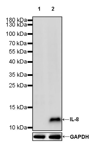
WB result of IL-8 Recombinnat Rabbit mAb
Primary antibody: IL-8 Rabbit mAb at 1/1000 dilution
Lane 1: untreated PC-3 whole cell lysate 20 µg
Lane 2: PC-3 treated with 2 µg/ml LPS and 500 ng/ml Brefeldin A for 5 hours whole cell lysate 20 µg
Secondary antibody: Goat Anti-rabbit IgG, (H+L), HRP conjugated at 1/10000 dilution
Predicted MW: 11 kDa
Observed MW: 12 kDa
IL-8 Rabbit mAb at 1/50 dilution (1 µg) immunoprecipitating IL-8 in 0.4 mg PC-3 treated with 2 µg/ml LPS and 500 ng/ml Brefeldin A for 5 hours whole cell lysate.
Western blot was performed on the immunoprecipitate using IL-8 Rabbit mAb at 1/1000 dilution.
Secondary antibody (HRP) for IP was used at 1/1000 dilution.
Lane 1: PC-3 treated with 2 µg/ml LPS and 500 ng/ml Brefeldin A for 5 hours whole cell lysate 20 µg (Input)
Lane 2: IL-8 Rabbit mAb IP in PC-3 treated with 2 µg/ml LPS and 500 ng/ml Brefeldin A for 5 hours whole cell lysate
Lane 3: Rabbit monoclonal IgG IP in PC-3 treated with 2 µg/ml LPS and 500 ng/ml Brefeldin A for 5 hours whole cell lysate
Predicted MW: 11 kDa
Observed MW: 12 kDa
This blot was developed with high sensitivity substrate
ICC analysis of PC-3 cells treated with LPS (2μg/ml, 5hr) + Brefeldin A (500ng/ml, 5hr) (top panel) and untreated PC-3 cells (below panel). Anti- IL-8 antibody was used at 1/500 dilution (Green) and incubated overnight at 4°C. Goat polyclonal Antibody to Rabbit IgG - H&L (Alexa Fluor® 488) was used as secondary antibody at 1/1000 dilution. The cells were fixed with 4% PFA and permeabilized with 0.1% PBS-Triton X-100. Nuclei were counterstained with DAPI (Blue). Counterstain with tubulin (Red).
 您现在的位置: