
PBS, 40% Glycerol, 0.05% BSA, 0.03% Proclin 300
12 months from date of receipt / reconstitution, -20 °C as supplied
| 应用 | 稀释度 |
|---|---|
| WB | 1:1000 |
| IHC-P | 1:1000 |
| ICC | 1:500 |
| IF | 1:200 |
| FCM | 1:2000 |
CD45RO is an important cell surface marker in immunology that belongs to the CD45 family (also known as Leukocyte Common Antigen, LCA). CD45 is a highly polymorphic transmembrane protein primarily expressed on the surface of white blood cells, including T cells, B cells, natural killer (NK) cells, monocytes, and granulocytes, and plays a crucial role in lymphocyte development, activation, and signaling. Specifically, CD45RO is an isoform of CD45 that is highly expressed on memory T cells but expressed at lower levels or not expressed on naive T cells. Memory T cells are T cells that have been activated and survived during previous immune responses, enabling them to rapidly and strongly activate upon re-encountering the same antigen, thus accelerating and enhancing immune responses. Therefore, CD45RO is often used to distinguish and identify memory T cells. It serves as an important marker in immune phenotyping analysis, disease diagnosis, immune response monitoring, and immunotherapy research.
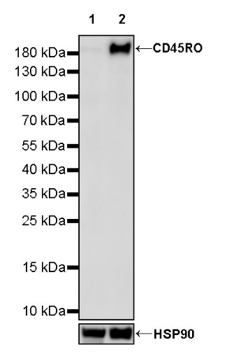
WB result of CD45RO Recombinant Mouse mAb
Primary antibody: CD45RO Mouse mAb at 1/1000 dilution
Lane 1: Jurkat whole cell lysate 20 µg
Lane 2: HuT 78 whole cell lysate 20 µg
Negative control: Jurkat whole cell lysate
Secondary antibody: Goat Anti-mouse IgG, (H+L), HRP conjugated at 1/10000 dilution
Predicted MW: 180 kDa
Observed MW: 180 kDa
Flow cytometric analysis of Jurkat (Human T cell leukemia T lymphocyte, left) / HuT 78 (Human Sezary syndrome cutaneous T lymphocyte, Right) labelling Human CD45RO antibody at 1/2000 dilution (0.1 μg) / (Red) compared with a Mouse monoclonal IgG (Black) isotype control and an unlabelled control (cells without incubation with primary antibody and secondary antibody) (Blue). Goat Anti - Mouse IgG Alexa Fluor® 488 was used as the secondary antibody.
Negative control: Jurkat
Flow cytometric analysis of human PBMC (human peripheral blood mononuclear cell) labelling CD45RO antibody at 1/2000 (0.1 μg) dilution (Right) compared with a Mouse monoclonal IgG isotype control (Left). Goat Anti - Mouse IgG Alexa Fluor® 488 was used as the secondary antibody. Then cells were stained with CD45RA - PE/Cy5.5 separately. Gated on viable lymphocytes.
IHC shows positive staining in paraffin-embedded human spleen. Anti-CD45RO antibody was used at 1/1000 dilution, followed by a HRP Polymer for Mouse & Rabbit IgG (ready to use). Counterstained with hematoxylin. Heat mediated antigen retrieval with Tris/EDTA buffer pH9.0 was performed before commencing with IHC staining protocol.
IHC shows positive staining in paraffin-embedded human tonsil. Anti-CD45RO antibody was used at 1/1000 dilution, followed by a HRP Polymer for Mouse & Rabbit IgG (ready to use). Counterstained with hematoxylin. Heat mediated antigen retrieval with Tris/EDTA buffer pH9.0 was performed before commencing with IHC staining protocol.
Negative control: IHC shows negative staining in paraffin-embedded human cerebral cortex. Anti-CD45RO antibody was used at 1/1000 dilution, followed by a HRP Polymer for Mouse & Rabbit IgG (ready to use). Counterstained with hematoxylin. Heat mediated antigen retrieval with Tris/EDTA buffer pH9.0 was performed before commencing with IHC staining protocol.
IHC shows positive staining in paraffin-embedded human thyroid cancer. Anti-CD45RO antibody was used at 1/1000 dilution, followed by a HRP Polymer for Mouse & Rabbit IgG (ready to use). Counterstained with hematoxylin. Heat mediated antigen retrieval with Tris/EDTA buffer pH9.0 was performed before commencing with IHC staining protocol.
IHC shows positive staining in paraffin-embedded non-Hodgkin’s T-cell lymphoma. Anti-CD45RO antibody was used at 1/1000 dilution, followed by a HRP Polymer for Mouse & Rabbit IgG (ready to use). Counterstained with hematoxylin. Heat mediated antigen retrieval with Tris/EDTA buffer pH9.0 was performed before commencing with IHC staining protocol.
IHC shows positive staining in paraffin-embedded human anaplastic large cell lymphoma. Anti-CD45RO antibody was used at 1/1000 dilution, followed by a HRP Polymer for Mouse & Rabbit IgG (ready to use). Counterstained with hematoxylin. Heat mediated antigen retrieval with Tris/EDTA buffer pH9.0 was performed before commencing with IHC staining protocol.
ICC shows positive staining in HuT 78 cells (top panel) and negative staining in Jurkat cells (below panel). Anti-CD45RO antibody was used at 1/500 dilution (Green) and incubated overnight at 4°C. Goat polyclonal Antibody to Rabbit IgG - H&L (Alexa Fluor® 488) was used as secondary antibody at 1/1000 dilution. The cells were fixed with 100% ice-cold methanol and permeabilized with 0.1% PBS-Triton X-100. Nuclei were counterstained with DAPI (Blue). Counterstain with tubulin (Red).
IF shows positive staining in paraffin-embedded human tonsil. Anti-CD45RO antibody was used at 1/200 dilution (Green) and incubated overnight at 4°C. Goat polyclonal Antibody to Mouse IgG - H&L (Alexa Fluor® 488) was used as secondary antibody at 1/1000 dilution. Counterstained with DAPI (Blue). Heat mediated antigen retrieval with EDTA buffer pH9.0 was performed before commencing with IF staining protocol.
IF shows positive staining in paraffin-embedded human anaplastic large cell lymphoma. Anti-CD45RO antibody was used at 1/200 dilution (Green) and incubated overnight at 4°C. Goat polyclonal Antibody to Mouse IgG - H&L (Alexa Fluor® 488) was used as secondary antibody at 1/1000 dilution. Counterstained with DAPI (Blue). Heat mediated antigen retrieval with EDTA buffer pH9.0 was performed before commencing with IF staining protocol.
 您现在的位置: