
12 months from date of receipt / reconstitution, -20 °C as supplied
| 应用 | 稀释度 |
|---|---|
| IHC-P | 1:2000 |
CXCR3, also known as C-X-C chemokine receptor type 3, is a G-protein-coupled receptor primarily expressed on the surface of activated T cells, B cells, and natural killer (NK) cells. It functions by binding to specific ligands on target cell membranes, inducing targeted migration and immune responses. CXCR3 plays a significant role in infection, autoimmune diseases, and tumor immunology. As a receptor for CXC chemokines such as CXCL9, CXCL10, and CXCL11, CXCR3 affects human mesangial cells (HMC) and other cell types through G-protein signaling. CXCR3 may also bind to CCL21, potentially contributing to chemotactic responses. Additionally, CXCR3 plays a crucial role in directing the migration of effector T cells to sites of inflammation and tumors, with its activation preferentially leading to high expression in Th1-type CD4+ T lymphocytes, effector CD8+ T lymphocytes, NK cells, and NKT cells. In recent years, numerous studies have confirmed that the expression of CXCR3 and its ligands in the body is closely related to tumor immunity, tumorigenesis, and metastasis. Therefore, CXCR3 has the potential to become a new indicator for assessing the prognosis of tumor patients and a new target for clinical tumor immunotherapy.
IHC shows positive staining in paraffin-embedded human tonsil. Anti-CXCR3 antibody was used at 1/2000 dilution, followed by a HRP Polymer for Mouse & Rabbit IgG (ready to use). Counterstained with hematoxylin. Heat mediated antigen retrieval with Tris/EDTA buffer pH9.0 was performed before commencing with IHC staining protocol.
IHC shows positive staining in paraffin-embedded human stomach. Anti-CXCR3 antibody was used at 1/2000 dilution, followed by a HRP Polymer for Mouse & Rabbit IgG (ready to use). Counterstained with hematoxylin. Heat mediated antigen retrieval with Tris/EDTA buffer pH9.0 was performed before commencing with IHC staining protocol.
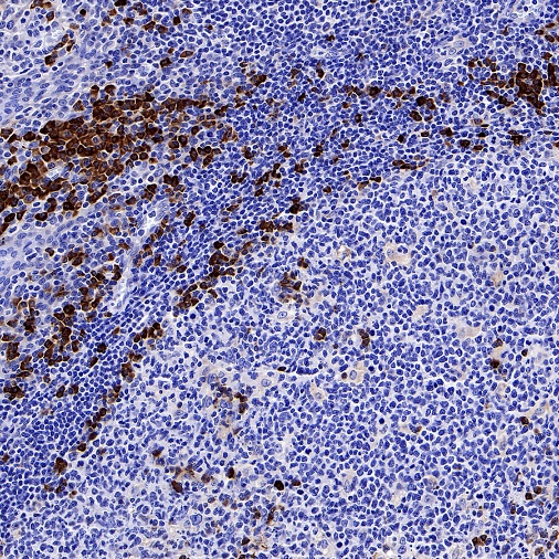
IHC shows positive staining in paraffin-embedded human Hodgkin’s lymphoma. Anti-CXCR3 antibody was used at 1/2000 dilution, followed by a HRP Polymer for Mouse & Rabbit IgG (ready to use). Counterstained with hematoxylin. Heat mediated antigen retrieval with Tris/EDTA buffer pH9.0 was performed before commencing with IHC staining protocol.
IHC shows positive staining in paraffin-embedded human cervical squamous cell carcinoma. Anti-CXCR3 antibody was used at 1/2000 dilution, followed by a HRP Polymer for Mouse & Rabbit IgG (ready to use). Counterstained with hematoxylin. Heat mediated antigen retrieval with Tris/EDTA buffer pH9.0 was performed before commencing with IHC staining protocol.
IHC shows positive staining in paraffin-embedded human colon cancer. Anti-CXCR3 antibody was used at 1/2000 dilution, followed by a HRP Polymer for Mouse & Rabbit IgG (ready to use). Counterstained with hematoxylin. Heat mediated antigen retrieval with Tris/EDTA buffer pH9.0 was performed before commencing with IHC staining protocol.
IHC shows positive staining in paraffin-embedded human pancreatic carcinoma. Anti-CXCR3 antibody was used at 1/2000 dilution, followed by a HRP Polymer for Mouse & Rabbit IgG (ready to use). Counterstained with hematoxylin. Heat mediated antigen retrieval with Tris/EDTA buffer pH9.0 was performed before commencing with IHC staining protocol.
 您现在的位置: