
12 months from date of receipt / reconstitution, 2 to 8 °C as supplied
| 应用 | 稀释度 |
|---|---|
| ICC | 1:500 |
| IF | 1:500 |
| ICFCM | 1:2000 |
Core component of nucleosome. Nucleosomes wrap and compact DNA into chromatin, limiting DNA accessibility to the cellular machineries which require DNA as a template. Histones thereby play a central role in transcription regulation, DNA repair, DNA replication and chromosomal stability. DNA accessibility is regulated via a complex set of post-translational modifications of histones, also called histone code, and nucleosome remodeling. H3.1 is enriched in PTMs associated with gene activation (K14 acetylation) and gene silencing (K9 dimethylation), suggesting that these mammalian H3 variants may, indeed, have separate biological functions.
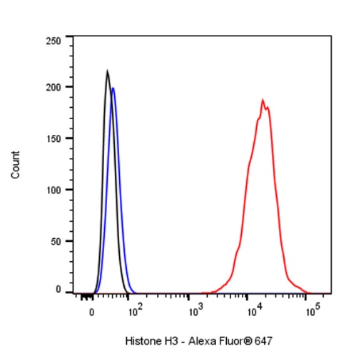
Flow cytometric analysis of 4% PFA fixed 90% methanol permeabilized A549 (Human lung carcinoma epithelial cell) cells labelling Histone H3 (Alexa Fluor® 647 Conjugate) antibody at 1/2000 dilution (0.1 μg) / (Red) compared with a Rabbit monoclonal IgG (Black) isotype control and an unlabelled control (cells without incubation with primary antibody and secondary antibody) (Blue).
Flow cytometric analysis of 4% PFA fixed 90% methanol permeabilized NIH/3T3 (Mouse embryonic fibroblast) cells labelling Histone H3 (Alexa Fluor® 647 Conjugate) antibody at 1/2000 dilution (0.1 μg) / (Red) compared with a Rabbit monoclonal IgG (Black) isotype control and an unlabelled control (cells without incubation with primary antibody and secondary antibody) (Blue).
Flow cytometric analysis of 4% PFA fixed 90% methanol permeabilized C6 (Rat glial tumor glial cell) cells labelling Histone H3 (Alexa Fluor® 647 Conjugate) antibody at 1/2000 dilution (0.1 μg) / (Red) compared with a Rabbit monoclonal IgG (Black) isotype control and an unlabelled control (cells without incubation with primary antibody and secondary antibody) (Blue).
ICC shows positive staining in HepG2 cells. Anti- Histone H3 (Alexa Fluor® 647 Conjugate) antibody was used at 1/500 dilution (magenta) and incubated overnight at 4°C. The cells were fixed with 100% ice-cold methanol and permeabilized with 0.1% PBS-Triton X-100. Nuclei were counterstained with DAPI (Blue).
ICC shows positive staining in NIH/3T3 cells. Anti- Histone H3 (Alexa Fluor® 647 Conjugate) antibody was used at 1/500 dilution (magenta) and incubated overnight at 4°C. The cells were fixed with 100% ice-cold methanol and permeabilized with 0.1% PBS-Triton X-100. Nuclei were counterstained with DAPI (Blue).
ICC shows positive staining in C6 cells. Anti- Histone H3 (Alexa Fluor® 647 Conjugate) antibody was used at 1/500 dilution (magenta) and incubated overnight at 4°C. The cells were fixed with 100% ice-cold methanol and permeabilized with 0.1% PBS-Triton X-100. Nuclei were counterstained with DAPI (Blue).
IF shows positive staining in paraffin-embedded human breast cancer. Anti- Histone H3 (Alexa Fluor® 647 Conjugate) antibody was used at 1/500 dilution (magenta) and incubated overnight at 4°C. Counterstained with DAPI (Blue). Heat mediated antigen retrieval with EDTA buffer pH9.0 was performed before commencing with IF staining protocol.
IF shows positive staining in paraffin-embedded human ovarian cancer. Anti- Histone H3 (Alexa Fluor® 647 Conjugate) antibody was used at 1/500 dilution (magenta) and incubated overnight at 4°C. Counterstained with DAPI (Blue). Heat mediated antigen retrieval with EDTA buffer pH9.0 was performed before commencing with IF staining protocol.
IF shows positive staining in paraffin-embedded mouse testis. Anti- Histone H3 (Alexa Fluor® 647 Conjugate) antibody was used at 1/500 dilution (magenta) and incubated overnight at 4°C. Counterstained with DAPI (Blue). Heat mediated antigen retrieval with EDTA buffer pH9.0 was performed before commencing with IF staining protocol.
IF shows positive staining in paraffin-embedded rat testis. Anti- Histone H3 (Alexa Fluor® 647 Conjugate) antibody was used at 1/500 dilution (magenta) and incubated overnight at 4°C. Counterstained with DAPI (Blue). Heat mediated antigen retrieval with EDTA buffer pH9.0 was performed before commencing with IF staining protocol.
 您现在的位置: