
12 months from date of receipt / reconstitution, -20°C as supplied
| 应用 | 稀释度 |
|---|---|
| WB | 1:1000 |
| ICC | 1:500 |
| ICFCM | 1:500 |
HMGB1 protein, or High Mobility Group Box 1 protein, is a highly conserved nuclear protein found widely in mammalian cells. HMGB1 is prevalent in various tissues such as lymphatic tissue, brain, liver, lung, heart, spleen, and kidney. In most tissues, it primarily resides in the nucleus, but in certain tissues like the liver and brain, it can also be found in the cytoplasm. Within the nucleus, HMGB1 interacts with ribosomes, transcription factors, and other components, participating in maintaining nucleosome structure and regulating gene expression. It can also modulate gene transcription by facilitating nucleosome sliding. Outside the cell, HMGB1 serves as a late-phase inflammatory mediator, involving inflammation, immune responses, tissue repair, and regeneration. It mediates these biological effects by interacting with various receptors, such as Toll-like receptor 4 and the receptor for advanced glycation end products. HMGB1 plays a crucial role in systemic inflammatory response syndrome, such as sepsis. Sepsis is a common complication of severe trauma, burns, and other diseases, and HMGB1 is a key inflammatory mediator in this condition.
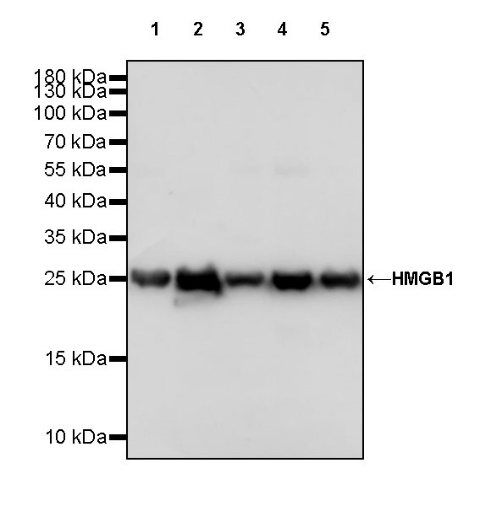
WB result of HMGB1 Rabbit pAb
Primary antibody: HMGB1 Rabbit pAb at 1/1000 dilution
Lane 1: HeLa whole cell lysate 20 µg
Lane 2: Jurkat whole cell lysate 20 µg
Lane 3: A431 whole cell lysate 20 µg
Lane 4: HepG2 whole cell lysate 20 µg
Lane 5: SK-BR-3 whole cell lysate 20 µg
Secondary antibody: Goat Anti-rabbit IgG, (H+L), HRP conjugated at 1/10000 dilution
Predicted MW: 25 kDa
Observed MW: 25 kDa
WB result of HMGB1 Rabbit pAb
Primary antibody: HMGB1 Rabbit pAb at 1/1000 dilution
Lane 1: NIH/3T3 whole cell lysate 20 µg
Secondary antibody: Goat Anti-rabbit IgG, (H+L), HRP conjugated at 1/10000 dilution
Predicted MW: 25 kDa
Observed MW: 25 kDa
WB result of HMGB1 Rabbit pAb
Primary antibody: HMGB1 Rabbit pAb at 1/1000 dilution
Lane 1: PC-12 whole cell lysate 20 µg
Secondary antibody: Goat Anti-rabbit IgG, (H+L), HRP conjugated at 1/10000 dilution
Predicted MW: 25 kDa
Observed MW: 25 kDa
Flow cytometric analysis of 4% PFA fixed 90% methanol permeabilized HeLa (Human cervix adenocarcinoma epithelial cell) labelling HMGB1 antibody at 1/500 dilution (0.1 μg)/ (Red) compared with a Rabbit monoclonal IgG (Black) isotype control and an unlabelled control (cells without incubation with primary antibody and secondary antibody) (Blue). Goat Anti - Rabbit IgG Alexa Fluor® 488 was used as the secondary antibody.
ICC shows positive staining in HeLa cells. Anti-HMGB1 antibody was used at 1/500 dilution (Green) and incubated overnight at 4°C. Goat polyclonal Antibody to Rabbit IgG - H&L (Alexa Fluor® 488) was used as secondary antibody at 1/1000 dilution. The cells were fixed with 4% PFA and permeabilized with 0.1% PBS-Triton X-100. Nuclei were counterstained with DAPI (Blue). Counterstain with tubulin (Red).
 您现在的位置: