
12 months from date of receipt / reconstitution, -20 °C as supplied
| 应用 | 稀释度 |
|---|---|
| WB | 1:1000 |
| IP | 1:50 |
IKKα, a member of the IKK family, possesses two kinase domains responsible for regulating the phosphorylation and degradation of IκB proteins. It specifically phosphorylates certain residues of IκB proteins (such as Ser32 and Ser36), leading to the degradation of IκB and subsequent activation of the NF-κB signaling pathway. IKKα plays a significant role in the immune system, particularly in regulating T-cell functions. It controls T-cell metabolism and proliferation, thereby influencing immune responses. Additionally, IKKα modulates the synthesis of cytokines, regulating interactions among immune cells. Studies have indicated abnormal expression or activity of IKKα in various cancers. Targeting IKKα represents a potential new strategy for cancer therapy. IKKα is also involved in the pathogenesis of multiple inflammatory diseases. Inhibitors of IKKα can alleviate inflammatory reactions, offering new avenues for treating these conditions.
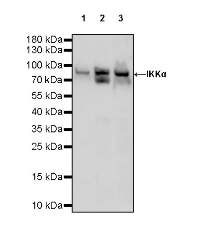
WB result of IKKα Rabbit pAb
Primary antibody: IKKα Rabbit pAb at 1/1000 dilution
Lane 1: HeLa whole cell lysate 20 µg
Lane 2: Jurkat whole cell lysate 20 µg
Lane 3: HCT 116 whole cell lysate 20 µg
Secondary antibody: Goat Anti-rabbit IgG, (H+L), HRP conjugated at 1/10000 dilution
Predicted MW: 85 kDa
Observed MW: 85 kDa
WB result of IKKα Rabbit pAb
Primary antibody: IKKα Rabbit pAb at 1/1000 dilution
Lane 1: RAW264.7 whole cell lysate 20 µg
Lane 2: mouse kidney lysate 20 µg
Secondary antibody: Goat Anti-rabbit IgG, (H+L), HRP conjugated at 1/10000 dilution
Predicted MW: 85 kDa
Observed MW: 85 kDa
WB result of IKKα Rabbit pAb
Primary antibody: IKKα Rabbit pAb at 1/1000 dilution
Lane 1: C6 whole cell lysate 20 µg
Secondary antibody: Goat Anti-rabbit IgG, (H+L), HRP conjugated at 1/10000 dilution
Predicted MW: 85 kDa
Observed MW: 85 kDa

IKKα Rabbit pAb at 1/50 dilution (1 µg) immunoprecipitating IKKα in 0.4 mg HCT116 whole cell lysate.
Western blot was performed on the immunoprecipitate using IKKα Rabbit pAb at 1/1000 dilution.
Secondary antibody (HRP) for IP was used at 1/1000 dilution.
Lane 1: HCT116 whole cell lysate 20 µg (Input)
Lane 2: IKKα Rabbit pAb IP in HCT116 whole cell lysate
Lane 3: Rabbit monoclonal IgG IP in HCT116 whole cell lysate
Predicted MW: 85 kDa
Observed MW: 85 kDa
 您现在的位置: