
12 months from date of receipt / reconstitution, -20 °C as supplied
| 应用 | 稀释度 |
|---|---|
| WB | 1:1000 |
| ICC | 1:500 |
| FCM | 1:500 |
CD272, also known as B and T lymphocyte attenuator (BTLA), is an antigen closely related to immune cell function. When the body is stimulated by antigenic substances, B-cells differentiate into plasma cells, producing immunoglobulins that can specifically bind to the corresponding antigens. CD272, as the B and T lymphocyte attenuator antigen, may play an important role in this process. In recent years, the application of CD272 in medical research has gradually increased. For example, in studies of acute myeloid leukemia, scientists have discovered that leukemia stem cells highly express CD272, and its high expression is negatively correlated with poor prognosis and survival rates of patients. This discovery provides a new target and research idea for the treatment of leukemia. By blocking the cross-linking signal between CD272 and its ligand, the growth of leukemia cells can be effectively inhibited, providing new possibilities for the treatment of leukemia.
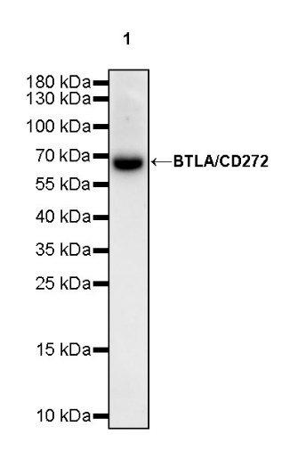
WB result of BTLA/CD272 Rabbit mAb
Primary antibody: BTLA/CD272 Rabbit mAb at 1/1000 dilution
Lane 1: mouse spleen lysate 20 µg
Secondary antibody: Goat Anti-rabbit IgG, (H+L), HRP conjugated at 1/10000 dilution
Predicted MW: 34 kDa
Observed MW: 68 kDa
(This blot was developed with high sensitivity substrate)
WB result of BTLA/CD272 Rabbit mAb
Primary antibody: BTLA/CD272 Rabbit mAb at 1/1000 dilution
Lane 1: rat spleen lysate 20 µg
Secondary antibody: Goat Anti-rabbit IgG, (H+L), HRP conjugated at 1/10000 dilution
Predicted MW: 34 kDa
Observed MW: 68 kDa
(This blot was developed with high sensitivity substrate)
Flow cytometric analysis of C57BL/6 mouse splenocytes labeling BTLA/CD272 at 1/500 (0.1 μg) dilution / (Right panel) compared with a Rabbit IgG, Isotype Control / (Left panel). Goat Anti -Rabbit IgG Alexa Fluor® 488 was used as the secondary antibody. Then cells were stained with CD3 - Alexa Fluor® 647 separately. Gated on total viable cells.
ICC shows positive staining in A20 cells. Anti- BTLA/CD272 antibody was used at 1/500 dilution (Green) and incubated overnight at 4°C. Goat polyclonal Antibody to Rabbit IgG - H&L (Alexa Fluor® 488) was used as secondary antibody at 1/1000 dilution. The cells were fixed with 4% PFA and permeabilized with 0.1% PBS-Triton X-100. Nuclei were counterstained with DAPI (Blue). Counterstain with tubulin (Red).
 您现在的位置: