
12 months from date of receipt / reconstitution, -20 °C as supplied
| 应用 | 稀释度 |
|---|---|
| WB | 1:1000 |
| IHC-P | 1:500 |
| ICC | 1:50 |
| ICFCM | 1:50 |
The CPS1 protein, also known as carbamoyl phosphate synthetase I, plays a crucial role in the urea cycle. The CPS1 protein has a molecular weight of 163kDa and is primarily located in the mitochondria of the liver. It is a key enzyme in the urea cycle, responsible for synthesizing urea from ammonia and CO2 in the body. This process is essential for eliminating excess ammonia in the body, making it crucial for ammonia detoxification in animals. Mutations in the CPS1 can lead to carbamoyl phosphate synthetase I deficiency (CPSID), a rare autosomal recessive genetic disorder that causes abnormalities in ammonia metabolism. This condition triggers a genetic metabolic disease characterized by hyperammonemia. Studies have shown that the CPS1 is closely related to various malignancies such as liver cancer, lung cancer, and colorectal cancer. It may affect the occurrence and development of tumors through multiple signal transduction pathways or by influencing pyrimidine synthesis.
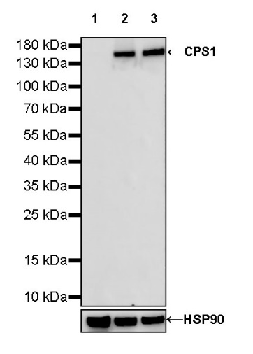
WB result of CPS1 Rabbit mAb
Primary antibody: CPS1 Rabbit mAb at 1/1000 dilution
Lane 1: U-2 OS whole cell lysate 20 µg
Lane 2: HeLa whole cell lysate 10 µg
Lane 3: SK-BR-3 whole cell lysate 20 µg
Negative control: U-2 OS whole cell lysate
Secondary antibody: Goat Anti-rabbit IgG, (H+L), HRP conjugated at 1/10000 dilution
Predicted MW: 165 kDa
Observed MW: 165 kDa
Flow cytometric analysis of 4% PFA fixed 90% methanol permeabilized HeLa (Human cervix adenocarcinoma epithelial cell) labelling CPS1 antibody at 1/50 dilution (1 μg)/ (Red) compared with a Rabbit monoclonal IgG (Black) isotype control and an unlabelled control (cells without incubation with primary antibody and secondary antibody) (Blue). Goat Anti - Rabbit IgG Alexa Fluor® 488 was used as the secondary antibody.
IHC shows positive staining in paraffin-embedded human liver. Anti-CPS1 antibody was used at 1/500 dilution, followed by a HRP Polymer for Mouse & Rabbit IgG (ready to use). Counterstained with hematoxylin. Heat mediated antigen retrieval with Tris/EDTA buffer pH9.0 was performed before commencing with IHC staining protocol.
IHC shows positive staining in paraffin-embedded human hepatocellular carcinoma. Anti-CPS1 antibody was used at 1/500 dilution, followed by a HRP Polymer for Mouse & Rabbit IgG (ready to use). Counterstained with hematoxylin. Heat mediated antigen retrieval with Tris/EDTA buffer pH9.0 was performed before commencing with IHC staining protocol.
ICC shows positive staining in HeLa cells. Anti-CPS1 antibody was used at 1/50 dilution (Green) and incubated overnight at 4°C. Goat polyclonal Antibody to Rabbit IgG - H&L (Alexa Fluor® 488) was used as secondary antibody at 1/1000 dilution. The cells were fixed with 100% ice-cold methanol and permeabilized with 0.1% PBS-Triton X-100. Nuclei were counterstained with DAPI (Blue). Counterstain with tubulin (Red).
 您现在的位置: