
PBS, 40% Glycerol, 0.05% BSA, 0.03% Proclin 300
12 months from date of receipt / reconstitution, -20 °C as supplied
| 应用 | 稀释度 |
|---|---|
| WB | 1:1000 |
| IHC-P | 1:500 |
| ICC | 1:500 |
| ICFCM | 1:500 |
NF-κB p105/p50 is a crucial component of the NF-κB (nuclear factor kappa-light-chain-enhancer of activated B cells) signaling pathway. The NF-κB family in mammals includes members such as RelA (p65), RelB, c-Rel, NF-κB1 (p50/p105), and NF-κB2 (p52/p100), which regulate gene expression through the formation of various homodimers or heterodimers. NF-κB p105 serves as the precursor form of NF-κB1 and primarily exists in this form within the cell. In response to specific stimuli, such as proinflammatory cytokines, free radicals, ultraviolet radiation, and bacterial or viral infections, NF-κB p105 is cleaved by the proteasome into the p50 subunit. This cleavage process liberates NF-κB from its complex with the inhibitory protein IκB, enabling its translocation to the nucleus and activation of target gene transcription. NF-κB p50 (as well as p52) only contains the Rel homology domain (RHD) and lacks the transactivation domain (TD). Therefore, p50/p52 homodimers cannot activate gene transcription and instead function as inhibitory molecules. However, when they form heterodimers with RelA, RelB, or c-Rel, which possess the TD, they are able to bind to DNA and activate transcription. NF-κB p105/p50 plays a pivotal role in regulating various physiological processes, including immune responses, inflammation, cell growth, and death. Abnormal expression or dysfunction of these proteins is closely associated with the development and progression of multiple diseases, such as cancer, inflammatory bowel disease, and rheumatoid arthritis.
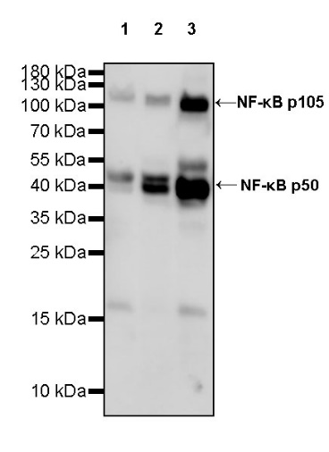
WB result of NF-κB p105/p50 Rabbit pAb
Primary antibody: NF-κB p105/p50 Rabbit pAb at 1/1000 dilution
Lane 1: HeLa whole cell lysate 20 µg
Lane 2: A431 whole cell lysate 20 µg
Lane 3: Jurkat whole cell lysate 20 µg
Secondary antibody: Goat Anti-rabbit IgG, (H+L), HRP conjugated at 1/10000 dilution
Predicted MW: 105 kDa
Observed MW: 50, 105 kDa
Exposure time: 180 s
WB result of NF-κB p105/p50 Rabbit pAb
Primary antibody: NF-κB p105/p50 Rabbit pAb at 1/1000 dilution
Lane 1: NIH/3T3 whole cell lysate 20 µg
Lane 2: mouse spleen lysate 20 µg
Secondary antibody: Goat Anti-rabbit IgG, (H+L), HRP conjugated at 1/10000 dilution
Predicted MW: 105 kDa
Observed MW: 50, 105 kDa
Exposure time: 180 s
WB result of NF-κB p105/p50 Rabbit pAb
Primary antibody: NF-κB p105/p50 Rabbit pAb at 1/1000 dilution
Lane 1: rat spleen lysate 20 µg
Secondary antibody: Goat Anti-rabbit IgG, (H+L), HRP conjugated at 1/10000 dilution
Predicted MW: 105 kDa
Observed MW: 50, 105 kDa
Exposure time: 180 s
Intracellular flow cytometric analysis of 4% PFA fixed 90% methanol permeabilized C2C12 (Mouse myoblasts myoblast), treated with 20ng/ml TNF-alpha for 30min (Red) or untreated (Green), labeling NF-κB p105/p50 at 1/500 dilution (0.1 μg) compared with a rabbit monoclonal IgG isotype control (Black) and an unlabeled control (cells without incubation with primary antibody and secondary antibody) (Blue). Goat Anti - Rabbit IgG Alexa Fluor® 488 was used as the secondary antibody.
IHC shows positive staining in paraffin-embedded human colon. Anti- NF-κB p105/p50 antibody was used at 1/500 dilution, followed by a HRP Polymer for Mouse & Rabbit IgG (ready to use). Counterstained with hematoxylin. Heat mediated antigen retrieval with Tris/EDTA buffer pH9.0 was performed before commencing with IHC staining protocol.
IHC shows positive staining in paraffin-embedded human tonsil. Anti- NF-κB p105/p50 antibody was used at 1/500 dilution, followed by a HRP Polymer for Mouse & Rabbit IgG (ready to use). Counterstained with hematoxylin. Heat mediated antigen retrieval with Tris/EDTA buffer pH9.0 was performed before commencing with IHC staining protocol.
IHC shows positive staining in paraffin-embedded human thyroid cancer. Anti- NF-κB p105/p50 antibody was used at 1/500 dilution, followed by a HRP Polymer for Mouse & Rabbit IgG (ready to use). Counterstained with hematoxylin. Heat mediated antigen retrieval with Tris/EDTA buffer pH9.0 was performed before commencing with IHC staining protocol.
IHC shows positive staining in paraffin-embedded mouse spleen. Anti- NF-κB p105/p50 antibody was used at 1/500 dilution, followed by a HRP Polymer for Mouse & Rabbit IgG (ready to use). Counterstained with hematoxylin. Heat mediated antigen retrieval with Tris/EDTA buffer pH9.0 was performed before commencing with IHC staining protocol.
IHC shows positive staining in paraffin-embedded rat spleen. Anti- NF-κB p105/p50 antibody was used at 1/500 dilution, followed by a HRP Polymer for Mouse & Rabbit IgG (ready to use). Counterstained with hematoxylin. Heat mediated antigen retrieval with Tris/EDTA buffer pH9.0 was performed before commencing with IHC staining protocol.
ICC analysis of C2C12 cells treated with TNF-alpha (20ng/ml,30min) (top panel) and C2C12 cells untreated with TNF-alpha (20ng/ml,30min (below panel). Anti-NF-κB p105/p50 antibody was used at 1/500 dilution (Green) and incubated overnight at 4°C. Goat polyclonal Antibody to Rabbit IgG - H&L (Alexa Fluor® 488) was used as secondary antibody at 1/1000 dilution. The cells were fixed with 4% PFA and permeabilized with 0.1% PBS-Triton X-100. Nuclei were counterstained with DAPI (Blue). Counterstain with tubulin (Red).
 您现在的位置: