
12 months from date of receipt / reconstitution, -20 °C as supplied
| 应用 | 稀释度 |
|---|---|
| IHC-P | 1:100 |
PD-L2, also known as Programmed Death-Ligand 2 or B7-DC, is a protein encoded by the PDCD1LG2 gene, which is located on chromosome 9p24.2. PD-L2 is one of the two Programmed Death Ligands (PD-L) and a member of the CD28 family of immune receptors. In the human body, PD-L2 is widely expressed in various tissues, including the placenta, heart, pancreas, lungs, and liver, but its expression is relatively low in the spleen, lymph nodes, and thymus. PD-L2 plays multiple roles in the immune system. Firstly, it can bind to PD-1 and inhibit the immune activation of T cells through downstream signaling pathways such as tyrosine phosphatase-2 (SHP-2). Secondly, PD-L2 can also bind to repulsive guidance molecule b (RGMb), activating the bone morphogenetic protein receptor pathway and promoting the functional activation of T cells. This dual functionality makes PD-L2 an important player in immune regulation. Furthermore, PD-L2 plays a significant role in tumor immune evasion mechanisms. It can both activate and inhibit T cells and may contribute to the development of immune-related pneumonia. Some studies have suggested that the expression of PD-L2 is associated with poor prognosis in esophageal cancer, supporting its role as a prognostic biomarker.
IHC shows positive staining in paraffin-embedded human tonsil. Anti- PD-L2 antibody was used at 1/100 dilution, followed by a HRP Polymer for Mouse & Rabbit IgG (ready to use). Counterstained with hematoxylin. Heat mediated antigen retrieval with Tris/EDTA buffer pH9.0 was performed before commencing with IHC staining protocol.
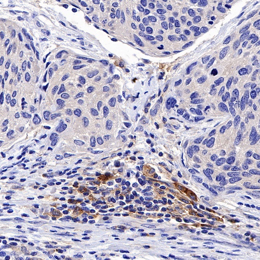
IHC shows positive staining in paraffin-embedded human cervical squamous cell carcinoma. Anti- PD-L2 antibody was used at 1/100 dilution, followed by a HRP Polymer for Mouse & Rabbit IgG (ready to use). Counterstained with hematoxylin. Heat mediated antigen retrieval with Tris/EDTA buffer pH9.0 was performed before commencing with IHC staining protocol.
IHC shows positive staining in paraffin-embedded human thyroid cancer. Anti- PD-L2 antibody was used at 1/100 dilution, followed by a HRP Polymer for Mouse & Rabbit IgG (ready to use). Counterstained with hematoxylin. Heat mediated antigen retrieval with Tris/EDTA buffer pH9.0 was performed before commencing with IHC staining protocol.
 您现在的位置: