
12 months from date of receipt / reconstitution, -20 °C as supplied
| 应用 | 稀释度 |
|---|---|
| WB | 1:1000 |
| IHC-P | 1:250 |
| ICC | 1:500 |
| ICFCM | 1:500 |
| IP | 1:50 |
FN14 protein, also known as fibroblast growth factor-inducible 14 or TWEAK receptor, is an I-type transmembrane protein receptor that belongs to the tumor necrosis factor receptor superfamily. This protein has a molecular weight of approximately 14 kDa and is encoded by the FN14 gene, which is located on chromosome 16p13. FN14 protein plays a crucial role in various biological processes, including cell growth, survival, and apoptosis. One significant function of FN14 protein is its role in tumor development and progression. In particular, studies have shown that FN14 is overexpressed in certain types of cancer, including non-small cell lung cancer (NSCLC) and hepatocellular carcinoma (HCC). In NSCLC, the overexpression of FN14 is associated with the activation of the epidermal growth factor receptor (EGFR), leading to tumor growth and metastasis. Similarly, in HCC, FN14 expression is linked to tumor progression and poor prognosis.
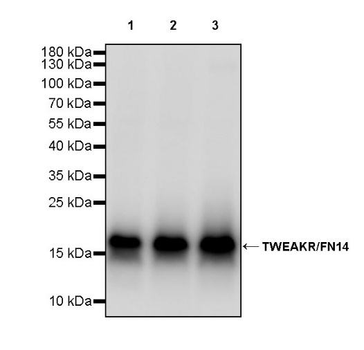
WB result of TWEAKR/FN14 Rabbit mAb
Primary antibody: TWEAKR/FN14 Rabbit mAb at 1/1000 dilution
Lane 1: HT-29 whole cell lysate 20 µg
Lane 2: HT-1080 whole cell lysate 20 µg
Lane 3: HUVEC whole cell lysate 20 µg
Secondary antibody: Goat Anti-rabbit IgG, (H+L), HRP conjugated at 1/10000 dilution
Predicted MW: 14 kDa
Observed MW: 17 kDa
WB result of TWEAKR/FN14 Rabbit mAb
Primary antibody: TWEAKR/FN14 Rabbit mAb at 1/1000 dilution
Lane 1: NIH/3T3 whole cell lysate 20 µg
Lane 2: C2C12 whole cell lysate 20 µg
Secondary antibody: Goat Anti-rabbit IgG, (H+L), HRP conjugated at 1/10000 dilution
Predicted MW: 14 kDa
Observed MW: 17 kDa
Flow cytometric analysis of 4% PFA fixed 90% methanol permeabilized HT-29 (Human colorectal adenocarcinoma epithelial cell) labelling TWEAKR/FN14 antibody at 1/500 dilution (0.1 μg)/ (Red) compared with a Rabbit monoclonal IgG (Black) isotype control and an unlabelled control (cells without incubation with primary antibody and secondary antibody) (Blue). Goat Anti - Rabbit IgG Alexa Fluor® 488 was used as the secondary antibody.

TWEAKR/FN14 Rabbit mAb at 1/50 dilution (1 µg) immunoprecipitating TWEAKR/FN14 in 0.4 mg HT-29 whole cell lysate.
Western blot was performed on the immunoprecipitate using TWEAKR/FN14 Rabbit mAb at 1/1000 dilution.
Secondary antibody (HRP) for IP was used at 1/1000 dilution.
Lane 1: HT-29 whole cell lysate 20 µg (Input)
Lane 2: TWEAKR/FN14 Rabbit mAb IP in HT-29 whole cell lysate
Lane 3: Rabbit monoclonal IgG IP in HT-29 whole cell lysate
Predicted MW: 14 kDa
Observed MW: 17 kDa
IHC shows positive staining in paraffin-embedded human cardiac muscle. Anti-TWEAKR/FN14 antibody was used at 1/250 dilution, followed by a HRP Polymer for Mouse & Rabbit IgG (ready to use). Counterstained with hematoxylin. Heat mediated antigen retrieval with Tris/EDTA buffer pH9.0 was performed before commencing with IHC staining protocol.
IHC shows positive staining in paraffin-embedded human ovarian cancer. Anti-TWEAKR/FN14 antibody was used at 1/250 dilution, followed by a HRP Polymer for Mouse & Rabbit IgG (ready to use). Counterstained with hematoxylin. Heat mediated antigen retrieval with Tris/EDTA buffer pH9.0 was performed before commencing with IHC staining protocol.
IHC shows positive staining in paraffin-embedded human pancreatic cancer. Anti-TWEAKR/FN14 antibody was used at 1/250 dilution, followed by a HRP Polymer for Mouse & Rabbit IgG (ready to use). Counterstained with hematoxylin. Heat mediated antigen retrieval with Tris/EDTA buffer pH9.0 was performed before commencing with IHC staining protocol.
IHC shows positive staining in paraffin-embedded human thyroid cancer. Anti-TWEAKR/FN14 antibody was used at 1/250 dilution, followed by a HRP Polymer for Mouse & Rabbit IgG (ready to use). Counterstained with hematoxylin. Heat mediated antigen retrieval with Tris/EDTA buffer pH9.0 was performed before commencing with IHC staining protocol.
ICC shows positive staining in HT-29 cells. Anti- TWEAK/FN14 antibody was used at 1/500 dilution (Green) and incubated overnight at 4°C. Goat polyclonal Antibody to Rabbit IgG - H&L (Alexa Fluor® 488) was used as secondary antibody at 1/1000 dilution. The cells were fixed with 4% PFA and permeabilized with 0.1% PBS-Triton X-100. Nuclei were counterstained with DAPI (Blue). Counterstain with tubulin (Red).
 您现在的位置: