
12 months from date of receipt / reconstitution, -20 °C as supplied
| 应用 | 稀释度 |
|---|---|
| WB | 1:1000 |
| IP | 1:50 |
| ICC | 1:500 |
| FCM | 1:5000 |
Epithelial cell adhesion molecule (EpCAM), also known as CD326 among other names, is a transmembrane glycoprotein mediating Ca2+-independent homotypic cell–cell adhesion in epithelia. EpCAM is also involved in cell signaling, migration, proliferation, and differentiation. Additionally, EpCAM has oncogenic potential via its capacity to upregulate c-myc, e-fabp, and cyclins A & E. Since EpCAM is expressed exclusively in epithelia and epithelial-derived neoplasms, EpCAM can be used as diagnostic marker for various cancers. It appears to play a role in tumorigenesis and metastasis of carcinomas, so it can also act as a potential prognostic marker and as a potential target for immunotherapeutic strategies.
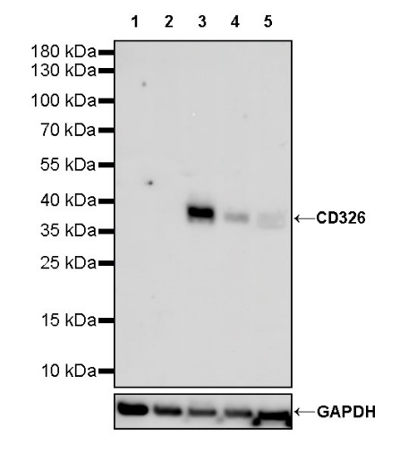
WB result of CD326 Rabbit mAb
Primary antibody: CD326 Rabbit mAb at 1/1000 dilution
Lane 1: HeLa whole cell lysate 20 µg
Lane 2: MB-MDA-231 whole cell lysate 20 µg
Lane 3: HT-29 whole cell lysate 20 µg
Lane 4: T-47D whole cell lysate 20 µg
Lane 5: MCF7 whole cell lysate 20 µg
Negative control: HeLa whole cell lysate; MB-MDA-231 whole cell lysate
Secondary antibody: Goat Anti-rabbit IgG, (H+L), HRP conjugated at 1/10000 dilution
Predicted MW: 35 kDa
Observed MW: 37 kDa
Flow cytometric analysis of Jurkat (Human T cell leukemia T lymphocyte, left) / HT29 (Human colorectal adenocarcinoma epithelial cell, Right) cells labelling CD326 antibody at 1/5000 dilution (0.01 μg) / (Red) compared with a Rabbit monoclonal IgG (Black) isotype control and an unlabelled control (cells without incubation with primary antibody and secondary antibody) (Blue). Goat Anti- Rabbit IgG Alexa Fluor® 488 was used as the secondary antibody.
Negative control: Jurkat
ICC shows positive staining in HT-29 cells (top panel) and negative staining in MDA-MB-231 cells (below panel). Anti-CD326 antibody was used at 1/500 dilution (Green) and incubated overnight at 4°C. Goat polyclonal Antibody to Rabbit IgG - H&L (Alexa Fluor® 488) was used as secondary antibody at 1/1000 dilution. The cells were fixed with 100% ice-cold methanol and permeabilized with 0.1% PBS-Triton X-100. Nuclei were counterstained with DAPI (Blue). Counterstain with tubulin (Red).
 您现在的位置: