
12 months from date of receipt / reconstitution, -20 °C as supplied
| 应用 | 稀释度 |
|---|---|
| WB | 1:1000 |
| IHC-P | 1:250 |
| ICC | 1:500 |
| ICFCM | 1:500 |
| IP | 1:50 |
Serine/threonine-protein phosphatase 2A, commonly known as Protein Phosphatase 2A (PP2A), is a highly conserved and ubiquitous Ser/Thr protein phosphatase found in eukaryotic organisms. It plays a pivotal role in various cellular processes such as cell cycle, metabolism, migration, and apoptosis. PP2A consists of a catalytic subunit C, a structural subunit A, and variable regulatory subunits B, with the catalytic subunit C (PP2A/C) serving as the core of its enzymatic activity and functionality. Specifically, PP2A regulates numerous cellular functions by catalyzing the dephosphorylation of proteins. Dephosphorylation is a crucial step in many cellular signaling pathways, affecting protein activity and function. Therefore, PP2A is instrumental in regulating cell proliferation, differentiation, apoptosis, and responses to external stimuli. Moreover, PP2A has close ties with the immune system. For instance, the activity of PP2A is significantly elevated in Treg cells (Regulatory T cells), and its high expression is essential for Treg cells to maintain their immunosuppressive function. This indicates that PP2A plays a crucial role in regulating immune responses and maintaining immune homeostasis.
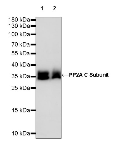
WB result of PP2A C Subunit Rabbit pAb
Primary antibody: PP2A C Subunit Rabbit pAb at 1/1000 dilution
Lane 1: HeLa whole cell lysate 20 µg
Lane 2: A431 whole cell lysate 20 µg
Secondary antibody: Goat Anti-rabbit IgG, (H+L), HRP conjugated at 1/10000 dilution
Predicted MW: 35 kDa
Observed MW: 36, 38 kDa
WB result of PP2A C Subunit Rabbit pAb
Primary antibody: PP2A C Subunit Rabbit pAb at 1/1000 dilution
Lane 1: NIH/3T3 whole cell lysate 20 µg
Secondary antibody: Goat Anti-rabbit IgG, (H+L), HRP conjugated at 1/10000 dilution
Predicted MW: 35 kDa
Observed MW: 36, 38 kDa
WB result of PP2A C Subunit Rabbit pAb
Primary antibody: PP2A C Subunit Rabbit pAb at 1/1000 dilution
Lane 1: C6 whole cell lysate 20 µg
Secondary antibody: Goat Anti-rabbit IgG, (H+L), HRP conjugated at 1/10000 dilution
Predicted MW: 35 kDa
Observed MW: 36, 38 kDa
Flow cytometric analysis of 4% PFA fixed 90% methanol permeabilized HeLa (Human cervix adenocarcinoma epithelial cell) labelling PP2A C Subunit antibody at 1/500 dilution (0.1 μg)/ (Red) compared with a Rabbit monoclonal IgG (Black) isotype control and an unlabelled control (cells without incubation with primary antibody and secondary antibody) (Blue). Goat Anti - Rabbit IgG Alexa Fluor® 488 was used as the secondary antibody.

PP2A C Subunit Rabbit pAb at 1/50 dilution (1 µg) immunoprecipitating PP2A C Subunit in 0.4 mg HeLa whole cell lysate.
Western blot was performed on the immunoprecipitate using PP2A C Subunit Rabbit pAb at 1/1000 dilution.
Secondary antibody (HRP) for IP was used at 1/1000 dilution.
Lane 1: HeLa whole cell lysate 20 µg (Input)
Lane 2: PP2A C Subunit Rabbit pAb IP in HeLa whole cell lysate
Lane 3: Rabbit monoclonal IgG IP in HeLa whole cell lysate
Predicted MW: 35 kDa
Observed MW: 36, 38 kDa
IHC shows positive staining in paraffin-embedded human kidney. Anti-PP2A C Subunit antibody was used at 1/250 dilution, followed by a HRP Polymer for Mouse & Rabbit IgG (ready to use). Counterstained with hematoxylin. Heat mediated antigen retrieval with Tris/EDTA buffer pH9.0 was performed before commencing with IHC staining protocol.
IHC shows positive staining in paraffin-embedded human breast cancer. Anti-PP2A C Subunit antibody was used at 1/250 dilution, followed by a HRP Polymer for Mouse & Rabbit IgG (ready to use). Counterstained with hematoxylin. Heat mediated antigen retrieval with Tris/EDTA buffer pH9.0 was performed before commencing with IHC staining protocol.
IHC shows positive staining in paraffin-embedded human lung squamous cell carcinoma. Anti-PP2A C Subunit antibody was used at 1/250 dilution, followed by a HRP Polymer for Mouse & Rabbit IgG (ready to use). Counterstained with hematoxylin. Heat mediated antigen retrieval with Tris/EDTA buffer pH9.0 was performed before commencing with IHC staining protocol.
IHC shows positive staining in paraffin-embedded mouse cerebral cortex. Anti-PP2A C Subunit antibody was used at 1/250 dilution, followed by a HRP Polymer for Mouse & Rabbit IgG (ready to use). Counterstained with hematoxylin. Heat mediated antigen retrieval with Tris/EDTA buffer pH9.0 was performed before commencing with IHC staining protocol.
ICC shows positive staining in HeLa cells. Anti- PP2A C Subunit antibody was used at 1/500 dilution (Green) and incubated overnight at 4°C. Goat polyclonal Antibody to Rabbit IgG - H&L (Alexa Fluor® 488) was used as secondary antibody at 1/1000 dilution. The cells were fixed with 4% PFA and permeabilized with 0.1% PBS-Triton X-100. Nuclei were counterstained with DAPI (Blue). Counterstain with tubulin (Red).
 您现在的位置: