
12 months from date of receipt / reconstitution, -20 °C as supplied
| 应用 | 稀释度 |
|---|---|
| IHC-P | 1:100-1:250 |
| ICC | 1:500 |
| ICFCM | 1:50 |
T-bet protein, also known as TBX21, is a transcription factor that regulates gene transcription, thereby influencing the development and function of immune cells. It plays a pivotal role in the immune system, particularly in regulating the differentiation and function of Th1 cells. As a Th1-specific transcription factor, T-bet induces the production of interferon-γ (IFN-γ), playing a decisive role in the differentiation of Th1 cells. It serves as a powerful transcriptional activator of the IFN-γ gene while also inducing and maintaining the expression of IL-12Rβ2. Additionally, T-bet can convert differentiating effector Th2 cells and fully differentiated Th2 cells into Th1 cells, accompanied by the production of large amounts of IFN-γ and the suppression of Th2-type cytokines such as IL-4, IL-5, and IL-13. During T-cell activation, T-bet expression is upregulated by IFN-γ. However, transforming growth factor-β (TGF-β) can suppress T-bet through a STAT1-dependent pathway, thereby inhibiting the differentiation of Th1 cells. Due to T-bet's crucial role in Th1 cell development and function, its activity is essential for maintaining normal immune system function. Th1 cells primarily participate in immune responses against intracellular pathogens, thus highlighting the significance of T-bet in protecting the body against such pathogens. However, abnormal TBX21 gene expression or function may be associated with immune system-related diseases, such as autoimmune disorders or immunodeficiencies.
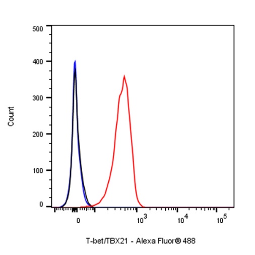
Flow cytometric analysis of 4% PFA fixed 90% methanol permeabilized NK-92 (Human malignant non-Hodgkin's lymphoma natural killer cell) labelling T-bet/TBX21 antibody at 1/50 dilution (1 μg)/ (Red) compared with a Rabbit monoclonal IgG (Black) isotype control and an unlabelled control (cells without incubation with primary antibody and secondary antibody) (Blue). Goat Anti - Rabbit IgG Alexa Fluor® 488 was used as the secondary antibody.
IHC shows positive staining in paraffin-embedded human spleen. Anti- T-bet/TBX21 antibody was used at 1/100 dilution, followed by a HRP Polymer for Mouse & Rabbit IgG (ready to use). Counterstained with hematoxylin. Heat mediated antigen retrieval with Tris/EDTA buffer pH9.0 was performed before commencing with IHC staining protocol.
IHC shows positive staining in paraffin-embedded human tonsil. Anti- T-bet/TBX21 antibody was used at 1/100 dilution, followed by a HRP Polymer for Mouse & Rabbit IgG (ready to use). Counterstained with hematoxylin. Heat mediated antigen retrieval with Tris/EDTA buffer pH9.0 was performed before commencing with IHC staining protocol.
Negative control: IHC shows negative staining in paraffin-embedded human cerebral cortex. Anti- T-bet/TBX21 antibody was used at 1/100 dilution, followed by a HRP Polymer for Mouse & Rabbit IgG (ready to use). Counterstained with hematoxylin. Heat mediated antigen retrieval with Tris/EDTA buffer pH9.0 was performed before commencing with IHC staining protocol.
IHC shows positive staining in paraffin-embedded human Hodgkin’s lymphoma. Anti- T-bet/TBX21 antibody was used at 1/100 dilution, followed by a HRP Polymer for Mouse & Rabbit IgG (ready to use). Counterstained with hematoxylin. Heat mediated antigen retrieval with Tris/EDTA buffer pH9.0 was performed before commencing with IHC staining protocol.
IHC shows positive staining in paraffin-embedded human thyroid cancer. Anti- T-bet/TBX21 antibody was used at 1/100 dilution, followed by a HRP Polymer for Mouse & Rabbit IgG (ready to use). Counterstained with hematoxylin. Heat mediated antigen retrieval with Tris/EDTA buffer pH9.0 was performed before commencing with IHC staining protocol.
IHC shows positive staining in paraffin-embedded mouse spleen. Anti- T-bet/TBX21 antibody was used at 1/250 dilution, followed by a HRP Polymer for Mouse & Rabbit IgG (ready to use). Counterstained with hematoxylin. Heat mediated antigen retrieval with Tris/EDTA buffer pH9.0 was performed before commencing with IHC staining protocol.
IHC shows positive staining in paraffin-embedded rat spleen. Anti- T-bet/TBX21 antibody was used at 1/250 dilution, followed by a HRP Polymer for Mouse & Rabbit IgG (ready to use). Counterstained with hematoxylin. Heat mediated antigen retrieval with Tris/EDTA buffer pH9.0 was performed before commencing with IHC staining protocol.
ICC shows positive staining in NK-92 cells. Anti-T-bet/TBX21 antibody was used at 1/500 dilution (Green) and incubated overnight at 4°C. Goat polyclonal Antibody to Rabbit IgG - H&L (Alexa Fluor® 488) was used as secondary antibody at 1/1000 dilution. The cells were fixed with 4% PFA and permeabilized with 0.1% PBS-Triton X-100. Nuclei were counterstained with DAPI (Blue). Counterstain with tubulin (Red).
 您现在的位置: