
| 应用 | 稀释度 |
|---|---|
| FCM | 5 μl per million cells in 100μl volume |
Aminopeptidase N (also called CD13) is located in the small-intestinal and renal microvillar membrane, and also in other plasma membranes. In the small intestine aminopeptidase N plays a role in the final digestion of peptides generated from hydrolysis of proteins by gastric and pancreatic proteases. Its function in proximal tubular epithelial cells and other cell types is less clear. The large extracellular carboxyterminal domain contains a pentapeptide consensus sequence characteristic of members of the zinc-binding metalloproteinase superfamily. CD13 is also used by some viruses as a receptor to which these viruses bind to and then enter cells. It is a receptor for human coronavirus 229E, feline coronavirus serotype II (FCoV-II), TGEV, PEDV, canine coronavirus genotype II (CCoV-II) as well as several Deltacoronaviruses.
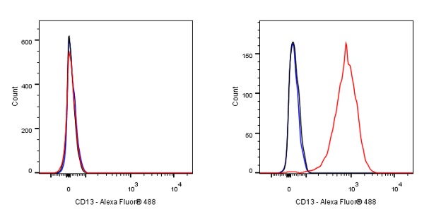
Flow cytometric analysis of CD13 expression on THP-1. Cells from the THP-1 (Human monocytic leukemia monocyte, Right) or HEK293 (Human embryonic kidney epithelial cell, Left) was stained with FITC Mouse IgG1, κ Isotype Control (Black line histogram) and SDT FITC Mouse Anti-Human CD13 antibody (Red line histogram) at 1 μg/test, cells without incubation with primary antibody and secondary antibody (Blue line histogram) was used as unlabelled control. Flow cytometry and data analysis were performed using BD FACSymphony™ A1 and FlowJo™ software.
 您现在的位置: