12 months from date of receipt / reconstitution, -20 °C as supplied
The CD19 antigen plays an important role in clinical oncology. In normal cells, it is the most ubiquitously expressed protein in the B lymphocyte lineage. CD19 expression is induced at the point of B lineage commitment during the differentiation of the hematopoietic stem cell, and its expression continues through preB and mature B cell differentiation until it is finally down-regulated during terminal differentiation into plasma cells. CD19 expression is maintained in B-lineage cells that have undergone neoplastic transformation, and therefore CD19 is useful in diagnosis of leukemias and lymphomas using monoclonal antibodies (mAbs) and flow cytometry [PMID: 8528044].
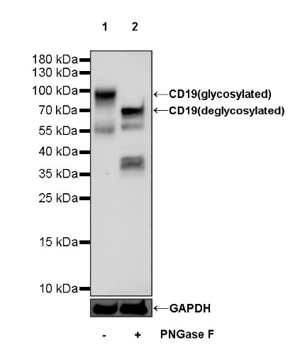
WB result of CD19 Mouse mAb
Primary antibody: CD19 Mouse mAb at 1/1000 dilution
Lane 1: untreated Raji whole cell lysate 20 µg
Lane 2: Raji treated with PNGase F whole cell lysate 20 µg
Secondary antibody: Goat Anti-mouse IgG, (H+L), HRP conjugated at 1/10000 dilution
Predicted MW: 61 kDa
Observed MW: 95 kDa
Flow cytometric analysis of human PBMC (human peripheral blood mononuclear cell) labelling CD19 antibody at 1/2000 (0.1 μg) dilution (Right) compared with a Mouse monoclonal IgG isotype control (Left). Goat Anti - Mouse IgG Alexa Fluor® 488 was used as the secondary antibody. Then cells were stained with CD3 - Alexa Fluor® 647 separately. Gated on total viable cells.
IHC shows positive staining in paraffin-embedded human spleen. Anti-CD19 antibody was used at 1/1000 dilution, followed by a HRP Polymer for Mouse & Rabbit IgG (ready to use). Counterstained with hematoxylin. Heat mediated antigen retrieval with Tris/EDTA buffer pH9.0 was performed before commencing with IHC staining protocol.
IHC shows positive staining in paraffin-embedded human tonsil. Anti-CD19 antibody was used at 1/500 dilution, followed by a HRP Polymer for Mouse & Rabbit IgG (ready to use). Counterstained with hematoxylin. Heat mediated antigen retrieval with Tris/EDTA buffer pH9.0 was performed before commencing with IHC staining protocol.
IHC shows positive staining in paraffin-embedded human diffuse large B-cell lymphoma. Anti-CD19 antibody was used at 1/500 dilution, followed by a HRP Polymer for Mouse & Rabbit IgG (ready to use). Counterstained with hematoxylin. Heat mediated antigen retrieval with Tris/EDTA buffer pH9.0 was performed before commencing with IHC staining protocol.
Negative control: IHC shows negative staining in paraffin-embedded human anaplastic large cell lymphoma. Anti-CD19 antibody was used at 1/500 dilution, followed by a HRP Polymer for Mouse & Rabbit IgG (ready to use). Counterstained with hematoxylin. Heat mediated antigen retrieval with Tris/EDTA buffer pH9.0 was performed before commencing with IHC staining protocol.
ICC shows positive staining in Raji cells (top panel) and negative staining in Jurkat cells (below panel). Anti-CD19 antibody was used at 1/500 dilution (Green) and incubated overnight at 4°C. Goat polyclonal Antibody to Mouse IgG - H&L (Alexa Fluor® 488) was used as secondary antibody at 1/1000 dilution. The cells were fixed with 100% ice-cold methanol and permeabilized with 0.1% PBS-Triton X-100. Nuclei were counterstained with DAPI (Blue). Counterstain with tubulin (Red).
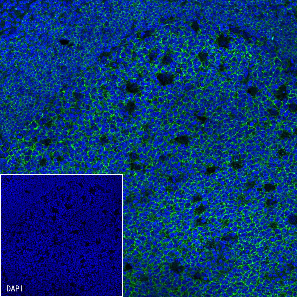
IF shows positive staining in paraffin-embedded human tonsil. Anti-CD19 antibody was used at 1/500 dilution (Green) and incubated overnight at 4°C. Goat polyclonal Antibody to Mouse IgG - H&L (Alexa Fluor® 488) was used as secondary antibody at 1/1000 dilution. Counterstained with DAPI (Blue). Heat mediated antigen retrieval with EDTA buffer pH9.0 was performed before commencing with IF staining protocol.
 您现在的位置: