
12 months from date of receipt / reconstitution, -20 °C as supplied
CD29 is an integrin component that mediates adhesion and involves in homing to sites of inflammation. It expresses in fibroblasts, platelets, T cells, monocytes, granulocytes(low), mast cells, endothelial cells and myoepithelium, also other diverse cell types. It does not express in red blood cells and spermatogonia. It is a myoepithelial marker, although established markers (SMA, CD10, p63, S100, maspin, calponin, GFAP, smooth muscle myosin) are more commonly used.
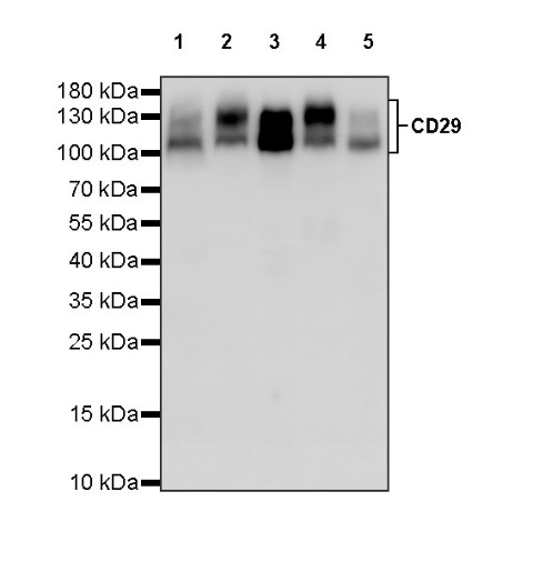
WB result of CD29 Mouse mAb
Primary antibody: CD29 Mouse mAb at 1/1000 dilution
Lane 1: HeLa whole cell lysate 20 µg
Lane 2: A-431 whole cell lysate 20 µg
Lane 3: A549 whole cell lysate 20 µg
Lane 4: U-87 MG whole cell lysate 20 µg
Lane 5: MCF7 whole cell lysate 20 µg
Secondary antibody: Goat Anti-mouse IgG, (H+L), HRP conjugated at 1/10000 dilution
Predicted MW: 88 kDa
Observed MW: 110~140 kDa
(This blot was developed with high sensitivity substrate)
Flow cytometric analysis of A549 (Human lung carcinoma epithelial cell) labelling CD29 antibody at 1/200 dilution (1 μg) / (Red) compared with a Mouse monoclonal IgG (Black) isotype control and an unlabelled control (cells without incubation with primary antibody and secondary antibody) (Blue). Goat Anti - Mouse IgG Alexa Fluor® 488 was used as the secondary antibody.
ICC shows positive staining in A549 cells. Anti-CD29 antibody was used at 1/100 dilution (Green) and incubated overnight at 4°C. Goat polyclonal Antibody to Mouse IgG - H&L (Alexa Fluor® 488) was used as secondary antibody at 1/1000 dilution. The cells were fixed with 100% ice-cold methanol and permeabilized with 0.1% PBS-Triton X-100. Nuclei were counterstained with DAPI (Blue).
 您现在的位置: