12 months from date of receipt / reconstitution, -20°C as supplied
| 应用 | 稀释度 |
|---|---|
| ICC | 1:500 |
| FCM | 1:200 |
CD69 protein is a type II transmembrane glycoprotein belonging to the C-type lectin receptor family and a member of the NK cell signaling transduction gene complex family. It is the earliest surface antigen expressed after the activation of T lymphocytes and can serve as a co-stimulatory signal to promote further activation and proliferation of T cells. Additionally, CD69 is also induced to express on NK cells, B cells, macrophages, neutrophils, and eosinophils. When external stimuli disappear, the expression of CD69 on activated T cells rapidly decreases. Furthermore, the expression of CD69 protein is associated with various disease states. For instance, it is closely related to the occurrence and development of malignancies, and its expression level in patients with chronic lymphocytic leukemia is tightly linked to factors such as the number of peripheral blood lymphocytes and clinical staging, indicating its potential as a new prognostic indicator for this disease. CD69 is also considered a marker of activation for eosinophils and certain other cells, and its high-level expression may suggest the presence of immune system diseases, hematological disorders, such as rheumatoid arthritis, systemic lupus erythematosus, leukemia, lymphoma, and more.
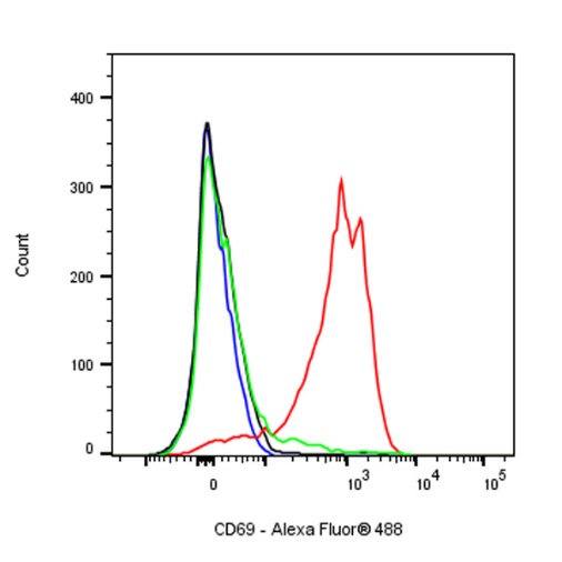
Flow cytometric analysis of C57BL/6 mouse splenocytes, treated overnight with 3μg/ml Concanavalin A (Red) or untreated (Green), labeling CD69 at 1/200 dilution (1 μg) compared with a Rat monoclonal IgG isotype control (Black) and an unlabeled control (cells without incubation with primary antibody and secondary antibody) (Blue). Goat Anti - Rat IgG Alexa Fluor® 488 was used as the secondary antibody.
ICC shows positive staining in Mouse splenocytes cells treated with Concanavalin A (3 µg/ml,overnight). Anti- CD69 antibody was used at 1/500 dilution (Green) and incubated overnight at 4°C. Goat polyclonal Antibody to Rat IgG - H&L (Alexa Fluor® 488) was used as secondary antibody at 1/1000 dilution. The cells were fixed with 4% PFA and permeabilized with 0.1% PBS-Triton X-100. Nuclei were counterstained with DAPI (Blue).
Negative control: ICC shows weak staining in Mouse splenocytes cells untreated with Concanavalin A (3 µg/ml, overnight). Anti- CD69 antibody was used at 1/500 dilution and incubated overnight at 4°C. Goat polyclonal Antibody to Rat IgG - H&L (Alexa Fluor® 488) was used as secondary antibody at 1/1000 dilution. The cells were fixed with 4% PFA and permeabilized with 0.1% PBS-Triton X-100. Nuclei were counterstained with DAPI (Blue).
 您现在的位置: