
PBS, 40% Glycerol, 0.05% BSA, 0.03% Proclin 300
12 months from date of receipt / reconstitution, -20 °C as supplied
| 应用 | 稀释度 |
|---|---|
| WB | 1:1000 |
| ICC | 1:500 |
| FCM | 1:500 |
| IP | 1:50 |
CD86 protein, also known as B7.2, is a crucial cell surface membrane protein belonging to the immunoglobulin superfamily with a molecular weight of approximately 80Kd. It is highly expressed on the surface of antigen-presenting cells. CD86 is expressed in various immune cells, including dendritic cells, monocytes, memory T lymphocytes, germinal center B lymphocytes, activated B lymphocytes, and activated T lymphocytes. CD86 protein plays a pivotal role in the immune system by interacting with CD28 and CTLA-4 (also known as CD152) proteins on the surface of T cells. The binding of CD86 to CD28 serves as a co-stimulatory signal for T cell activation, while the interaction between CD86 and CTLA-4 negatively regulates T cell activation and reduces immune responses. Additionally, CD86 protein contains two extracellular immunoglobulin domains (IgV and IgC), which bind to intercellular adhesion molecule 1 (ICAM-1) and participate in immune cell contact and signal transduction. Abnormal expression of CD86 is closely associated with the occurrence and development of various immune-related diseases. Therefore, understanding the structure and function of CD86 protein is crucial for revealing its immune regulatory mechanisms and providing new theoretical bases for disease treatment. Furthermore, experimental studies have demonstrated that CD86 protein plays a significant role in the activation and differentiation of immune cells, holding potential therapeutic value in immune-related diseases and offering a new breakthrough in the study of immune regulatory mechanisms.
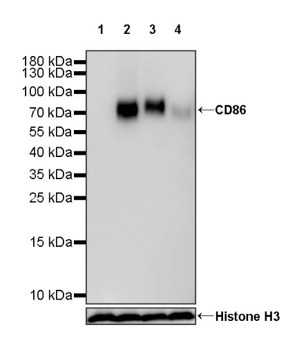
WB result of CD86 Rabbit mAb
Primary antibody: CD86 Rabbit mAb at 1/1000 dilution
Lane 1: Jurkat whole cell lysate 20 µg
Lane 2: Raji whole cell lysate 20 µg
Lane 3: Daudi whole cell lysate 20 µg
Lane 4: Ramos whole cell lysate 20 µg
Negative control: Jurkat whole cell lysate
Secondary antibody: Goat Anti-rabbit IgG, (H+L), HRP conjugated at 1/10000 dilution
Predicted MW: 37 kDa
Observed MW: 70~90 kDa
Flow cytometric analysis of Jurkat (Human T cell leukemia T lymphocyte, left) / Raji (Human Burkitt's lymphoma B lymphocyte, Right) cells labelling CD86 antibody at 1/500 dilution (0.1 μg) / (Red) compared with a Rabbit monoclonal IgG (Black) isotype control and an unlabelled control (cells without incubation with primary antibody and secondary antibody) (Blue). Goat Anti - Rabbit IgG Alexa Fluor® 488 was used as the secondary antibody.
Negative control: Jurkat
Flow cytometric analysis of human PBMC (human peripheral blood mononuclear cell) labelling CD86 antibody at 1/500 (0.1 μg) dilution (Right) compared with a Rabbit monoclonal IgG isotype control (Left). Goat Anti - Rabbit IgG Alexa Fluor® 488 was used as the secondary antibody. Then cells were stained with CD14 - Alexa Fluor® 647 separately. Gated on total viable cells.
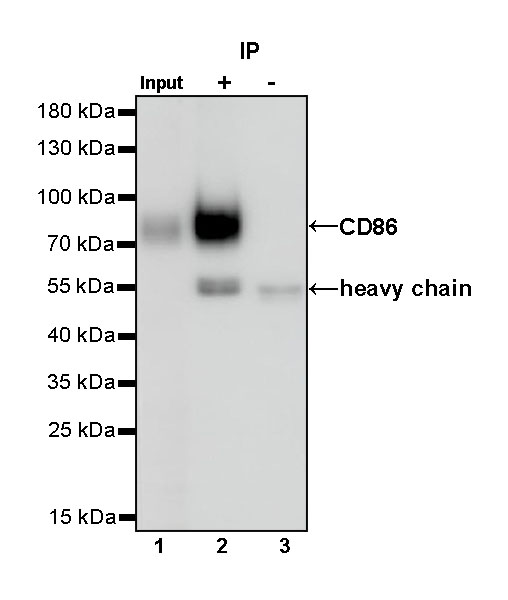
CD86 Rabbit mAb at 1/50 dilution (1 µg) immunoprecipitating CD86 in 0.4 mg Daudi whole cell lysate.
Western blot was performed on the immunoprecipitate using CD86 Rabbit mAb at 1/1000 dilution.
Secondary antibody (HRP) for IP was used at 1/1000 dilution.
Lane 1: Daudi whole cell lysate 20 µg (Input)
Lane 2: CD86 Rabbit mAb IP in Daudi whole cell lysate
Lane 3: Rabbit monoclonal IgG IP in Daudi whole cell lysate
Predicted MW: 37 kDa
Observed MW: 70~90 kDa
ICC shows positive staining in Raji cells (top panel) and negative staining in Jurkat cells (below panel). Anti-CD86 antibody was used at 1/500 dilution (Green) and incubated overnight at 4°C. Goat polyclonal Antibody to Rabbit IgG - H&L (Alexa Fluor® 488) was used as secondary antibody at 1/1000 dilution. The cells were fixed with 4% PFA and permeabilized with 0.1% PBS-Triton X-100. Nuclei were counterstained with DAPI (Blue). Counterstain with tubulin (Red).
 您现在的位置: