
PBS, 40% Glycerol, 0.05% BSA, 0.03% Proclin 300
12 months from date of receipt / reconstitution, -20 °C as supplied
Isotype control antibodies, to estimate the nonspecific binding of target. Use at concentrations comparable to those of the specific antibody of interest.
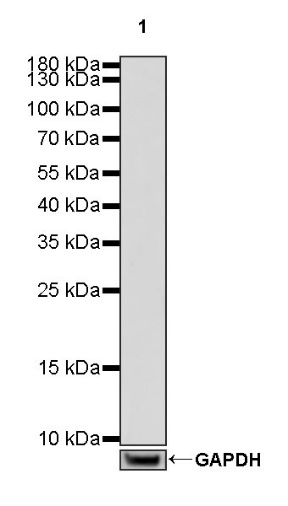
WB result of Mouse mAb IgG2b, κ Isotype Control
Primary antibody: Mouse mAb IgG2b, κ Isotype Control at 1/100 dilution
Lane 1: THP-1 whole cell lysate 20 µg
Secondary antibody: Goat Anti-mouse IgG, (H+L), HRP conjugated at 1/10000 dilution
Flow cytometric analysis of Hut 78 (Human Sezary syndrome cutaneous T lymphocyte) labeling Mouse mAb IgG2b, κ Isotype Control at 1/20 (1 μg) dilution (Left) / (Red) compared with CD6 antibody at 1/200 (1 μg) dilution (S0B0379) (Right) / (Red), Mouse monoclonal IgG isotype control (Left)/(Black) compared with Mouse mAb IgG2b, κ Isotype Control (Right) / (Black) and an unlabelled control (cells without incubation with primary antibody and secondary antibody) (Blue). Goat Anti - Mouse IgG Alexa Fluor® 488 was used as the secondary antibody.
Flow cytometric analysis of 4% paraformaldehyde fixed 90% methanol permeabilized HeLa (human cervical adenocarcinoma epithelial cell) labelling Mouse mAb IgG2b, κ Isotype Control at 1/20 (1 μg) dilution (Left) / (Red) compared with α-tubulin antibody (Right) / (Red), Mouse monoclonal IgG isotype control (Left) / (Black) compared with Mouse mAb IgG2b, κ Isotype Control (Right) / (Black) and an unlabelled control (cells without incubation with primary antibody and secondary antibody) (Blue). Goat Anti - Mouse IgG Alexa Fluor® 488 was used as the secondary antibody.
Flow cytometric analysis of 4% paraformaldehyde fixed 90% methanol permeabilized NIH/3T3 (Mouse embryonic fibroblast) labelling Mouse mAb IgG2b, κ Isotype Control at 1/20 (1 μg) dilution (Left) / (Red) compared with α-tubulin antibody (Right) / (Red), Mouse monoclonal IgG isotype control (Left) / (Black) compared with Mouse mAb IgG2b, κ Isotype Control (Right) / (Black) and an unlabelled control (cells without incubation with primary antibody and secondary antibody) (Blue). Goat Anti - Mouse IgG Alexa Fluor® 488 was used as the secondary antibody.
IHC shows negative staining in paraffin-embedded human tonsil. Mouse mAb IgG2b, κ Isotype Control was used at 1/50 dilution, followed by a HRP Polymer for Mouse & Rabbit IgG (ready to use). Counterstained with hematoxylin. Heat mediated antigen retrieval with Tris/EDTA buffer pH9.0 was performed before commencing with IHC staining protocol.
IHC shows negative staining in paraffin-embedded rat spleen. Mouse mAb IgG2b, κ Isotype Control was used at 1/50 dilution, followed by a HRP Polymer for Mouse & Rabbit IgG (ready to use). Counterstained with hematoxylin. Heat mediated antigen retrieval with Tris/EDTA buffer pH9.0 was performed before commencing with IHC staining protocol.
 您现在的位置: