
12 months from date of receipt / reconstitution, -20 °C as supplied
| 应用 | 稀释度 |
|---|---|
| WB | 1:1000 |
| ICC | 1:500 |
| ICFCM | 1:5000 |
| IP | 1:50 |
Runx1 is a crucial transcription factor that binds to the core elements of many enhancers and promoters, regulating the expression of specific genes. This protein plays a pivotal role in cellular development, particularly in the development of hematopoietic stem cells. RUNX1 interacts with CBFβ (core-binding factor β) to form a set of transcription factors, and this interaction is essential in the development of neurons and hematopoietic stem cells. However, when CBFβ undergoes translocation, it can form a fusion protein CBFβ-SMMHC that abnormally binds tightly to RUNX1, participating in the pathogenesis of acute lymphocytic leukemia (ALL). Therefore, the RUNX1−CBFβ interaction is also considered an important target for cancer treatment. Furthermore, research on RUNX1 protein has revealed its association with a range of cancers, particularly leukemia. Chromosome translocations involving the RUNX1 gene have been clearly linked to several types of leukemia.
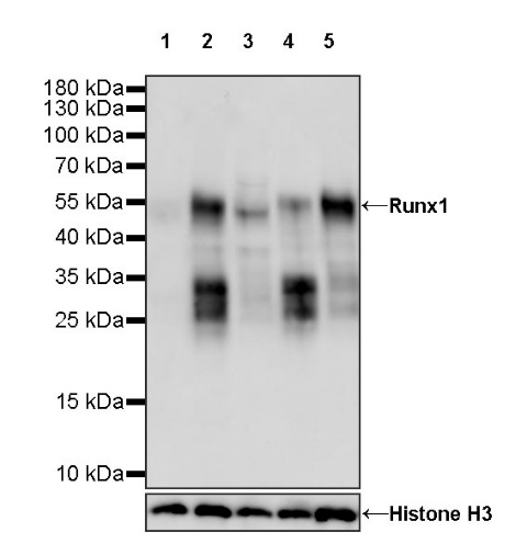
WB result of Runx1 Rabbit mAb
Primary antibody: Runx1 Rabbit mAb at 1/1000 dilution
Lane 1: HEK-293 whole cell lysate 20 µg
Lane 2: Jurkat whole cell lysate 20 µg
Lane 3: THP-1 whole cell lysate 20 µg
Lane 4: MOLT-4 whole cell lysate 20 µg
Lane 5: SW620 whole cell lysate 20 µg
Weak expression: HEK-293 whole cell lysate
Secondary antibody: Goat Anti-rabbit IgG, (H+L), HRP conjugated at 1/10000 dilution
Predicted MW: 48 kDa
Observed MW: 55 kDa
WB result of Runx1 Rabbit mAb
Primary antibody: Runx1 Rabbit mAb at 1/1000 dilution
Lane 1: mouse thymus lysate 20 µg
Secondary antibody: Goat Anti-rabbit IgG, (H+L), HRP conjugated at 1/10000 dilution
Predicted MW: 48 kDa
Observed MW: 55 kDa
WB result of Runx1 Rabbit mAb
Primary antibody: Runx1 Rabbit mAb at 1/1000 dilution
Lane 1: rat thymus lysate 20 µg
Secondary antibody: Goat Anti-rabbit IgG, (H+L), HRP conjugated at 1/10000 dilution
Predicted MW: 48 kDa
Observed MW: 55 kDa
Flow cytometric analysis of 4% PFA fixed 90% methanol permeabilized Jurkat (Human T cell leukemia T lymphocyte) labelling Runx1 antibody at 1/5000 dilution (0.01 μg) / (Red) compared with a Rabbit monoclonal IgG (Black) isotype control and an unlabelled control (cells without incubation with primary antibody and secondary antibody) (Blue). Goat Anti - Rabbit IgG Alexa Fluor® 488 was used as the secondary antibody.
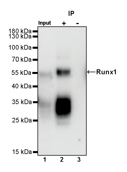
Runx1 Rabbit mAb at 1/50 dilution (1 µg) immunoprecipitating Runx1 in 0.4 mg Jurkat whole cell lysate.
Western blot was performed on the immunoprecipitate using Runx1 Rabbit mAb at 1/1000 dilution.
Secondary antibody (HRP) for IP was used at 1/1000 dilution.
Lane 1: Jurkat whole cell lysate 10 µg (Input)
Lane 2: Runx1 Rabbit mAb IP in Jurkat whole cell lysate
Lane 3: Rabbit monoclonal IgG IP in Jurkat whole cell lysate
Predicted MW: 48 kDa
Observed MW: 55 kDa
This blot was developed with high sensitivity substrate
ICC shows positive staining in Jurkat cells (top panel) and weak staining in HEK293 cells (below panel). Anti-RUNX1 antibody was used at 1/500 dilution (Green) and incubated overnight at 4°C. Goat polyclonal Antibody to Rabbit IgG - H&L (Alexa Fluor® 488) was used as secondary antibody at 1/1000 dilution. The cells were fixed with 100% ice-cold methanol and permeabilized with 0.1% PBS-Triton X-100. Nuclei were counterstained with DAPI (Blue). Counterstain with tubulin (Red).
 您现在的位置: