
12 months from date of receipt / reconstitution, -20 °C as supplied
| 应用 | 稀释度 |
|---|---|
| WB | 1:1000 |
| IP | 1:50 |
| ICC | 1:500 |
| ICFCM | 1:50 |
ADAM10 is a sheddase, and has a broad specificity for peptide hydrolysis reactions. ADAM10 cleaves ephrin, within the ephrin/eph complex, formed between two cell surfaces. When ephrin is freed from the opposing cell, the entire ephrin/eph complex is endocytosed. In neurons, ADAM10 is the most important enzyme, with α-secretase activity for proteolytic processing of the amyloid precursor protein. ADAM10, along with ADAM17, cleaves the ectodomain of the triggering receptor expressed on myeloid cells 2 (TREM2), to produce soluble TREM2 (sTREM2), which has been proposed as a CSF and sera biomarker of neurodegeneration. ADAM10 being a major determinant of HER2 shedding, the inhibition of which, may provide a novel therapeutic approach for treating breast cancer and a variety of other cancers with active HER2 signaling.
WB result of ADAM10 Rabbit pAb
Primary antibody: ADAM10 Rabbit pAb at 1/1000 dilution
Lane 1: A-431 whole cell lysate 20 µg
Lane 2: SK-OV-3 whole cell lysate 20 µg
Secondary antibody: Goat Anti-rabbit IgG, (H+L), HRP conjugated at 1/10000 dilution
Predicted MW: 84 kDa
Observed MW: 68, 90 kDa
(This blot was developed with high sensitivity substrate)
WB result of ADAM10 Rabbit pAb
Primary antibody: ADAM10 Rabbit pAb at 1/1000 dilution
Lane 1: RAW 264.7 whole cell lysate 20 µg
Secondary antibody: Goat Anti-rabbit IgG, (H+L), HRP conjugated at 1/10000 dilution
Predicted MW: 84 kDa
Observed MW: 68, 90 kDa
(This blot was developed with high sensitivity substrate)
Flow cytometric analysis of 4% PFA fixed 90% methanol permeabilized A431 (Human epidermoid carcinoma epithelial cell) labelling ADAM10 antibody at 1/50 dilution (1 μg) / (Red) compared with a Rabbit monoclonal IgG (Black) isotype control and an unlabelled control (cells without incubation with primary antibody and secondary antibody) (Blue). Goat Anti - Rabbit IgG Alexa Fluor® 488 was used as the secondary antibody.
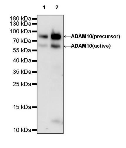
ADAM10 Rabbit mAb at 1/50 dilution (1 µg) immunoprecipitating ADAM10 in 0.4 mg A431 whole cell lysate.
Western blot was performed on the immunoprecipitate using ADAM10 Rabbit mAb at 1/1000 dilution.
Secondary antibody (HRP) for IP was used at 1/400 dilution.
Lane 1: A431 whole cell lysate 20 µg (Input)
Lane 2: ADAM10 Rabbit mAb IP in A431 whole cell lysate
Lane 3: Rabbit monoclonal IgG IP in A431 whole cell lysate
Predicted MW: 84 kDa
Observed MW: 68, 90 kDa
ICC shows positive staining in A431 cells. Anti-ADAM10 antibody was used at 1/500 dilution (Green) and incubated overnight at 4°C. Goat polyclonal Antibody to Rabbit IgG - H&L (Alexa Fluor® 488) was used as secondary antibody at 1/1000 dilution. The cells were fixed with 4% PFA and permeabilized with 0.1% PBS-Triton X-100. Nuclei were counterstained with DAPI (Blue). Counterstain with tubulin (Red).
 您现在的位置: