
12 months from date of receipt / reconstitution, -20 °C as supplied
| 应用 | 稀释度 |
|---|---|
| WB | 1:1000 |
| IHC-P | 1:2000 |
| ICC | 1:500 |
As an isocitrate dehydrogenase, IDH1 catalyzes the reversible oxidative decarboxylation of isocitrate to yield α-ketoglutarate (α-KG) as part of the TCA cycle in glucose metabolism. This step also allows for the concomitant reduction of nicotinamide adenine dinucleotide phosphate (NADP+) to reduced nicotinamide adenine dinucleotide phosphate (NADPH). Since NADPH and α-KG function in cellular detoxification processes in response to oxidative stress, IDH1 also indirectly participates in mitigating oxidative damage. In addition, IDH1 is key to β-oxidation of unsaturated fatty acids in the peroxisomes of liver cells. IDH1 also participates in the regulation of glucose-induced insulin secretion. Notably, IDH1 is the primary producer of NADPH in most tissues, especially in brain. Within cells, IDH1 has been observed to localize to the cytoplasm, peroxisome, and endoplasmic reticulum. Mutations in IDH1 have been shown to cause metaphyseal chondromatosis with aciduria. Mutations in IDH1 are also implicated in cancer.
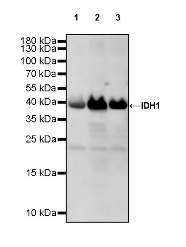
WB result of IDH1 Rabbit mAb
Primary antibody: IDH1 Rabbit mAb at 1/1000 dilution
Lane 1: 293T whole cell lysate 20 µg
Lane 2: Hep G2 whole cell lysate 20 µg
Lane 3: HeLa whole cell lysate 20 µg
Secondary antibody: Goat Anti-rabbit IgG, (H+L), HRP conjugated at 1/10000 dilution
Predicted MW: 46 kDa
Observed MW: 40 kDa
WB result of IDH1 Rabbit mAb
Primary antibody: IDH1 Rabbit mAb at 1/1000 dilution
Lane 1: Neuro-2a whole cell lysate 20 µg
Lane 2: C2C12 whole cell lysate 20 µg
Secondary antibody: Goat Anti-rabbit IgG, (H+L), HRP conjugated at 1/10000 dilution
Predicted MW: 46 kDa
Observed MW: 40 kDa
WB result of IDH1 Rabbit mAb
Primary antibody: IDH1 Rabbit mAb at 1/1000 dilution
Lane 1: rat skeletal muscle lysate 20 µg
Secondary antibody: Goat Anti-rabbit IgG, (H+L), HRP conjugated at 1/10000 dilution
Predicted MW: 46 kDa
Observed MW: 40 kDa
IHC shows positive staining in paraffin-embedded human kidney. Anti-IDH1 antibody was used at 1/2000 dilution, followed by a HRP Polymer for Mouse & Rabbit IgG (ready to use). Counterstained with hematoxylin. Heat mediated antigen retrieval with Tris/EDTA buffer pH9.0 was performed before commencing with IHC staining protocol.
IHC shows positive staining in paraffin-embedded human prostate. Anti-IDH1 antibody was used at 1/2000 dilution, followed by a HRP Polymer for Mouse & Rabbit IgG (ready to use). Counterstained with hematoxylin. Heat mediated antigen retrieval with Tris/EDTA buffer pH9.0 was performed before commencing with IHC staining protocol.
Negative control: IHC shows negative staining in paraffin-embedded human skeletal muscle. Anti-IDH1 antibody was used at 1/2000 dilution, followed by a HRP Polymer for Mouse & Rabbit IgG (ready to use). Counterstained with hematoxylin. Heat mediated antigen retrieval with Tris/EDTA buffer pH9.0 was performed before commencing with IHC staining protocol.
IHC shows positive staining in paraffin-embedded human colon cancer. Anti-IDH1 antibody was used at 1/2000 dilution, followed by a HRP Polymer for Mouse & Rabbit IgG (ready to use). Counterstained with hematoxylin. Heat mediated antigen retrieval with Tris/EDTA buffer pH9.0 was performed before commencing with IHC staining protocol.
IHC shows positive staining in paraffin-embedded human endometrial cancer. Anti-IDH1 antibody was used at 1/2000 dilution, followed by a HRP Polymer for Mouse & Rabbit IgG (ready to use). Counterstained with hematoxylin. Heat mediated antigen retrieval with Tris/EDTA buffer pH9.0 was performed before commencing with IHC staining protocol.
IHC shows positive staining in paraffin-embedded mouse kidney. Anti-IDH1 antibody was used at 1/2000 dilution, followed by a HRP Polymer for Mouse & Rabbit IgG (ready to use). Counterstained with hematoxylin. Heat mediated antigen retrieval with Tris/EDTA buffer pH9.0 was performed before commencing with IHC staining protocol.
ICC shows positive staining in 293T cells. Anti-IDH1 antibody was used at 1/500 dilution (Green) and incubated overnight at 4°C. Goat polyclonal Antibody to Rabbit IgG - H&L (Alexa Fluor® 488) was used as secondary antibody at 1/1000 dilution. The cells were fixed with 4% PFA and permeabilized with 0.1% PBS-Triton X-100. Nuclei were counterstained with DAPI (Blue). Counterstain with tubulin (Red).
ICC shows positive staining in HeLa cells. Anti-IDH1 antibody was used at 1/500 dilution (Green) and incubated overnight at 4°C. Goat polyclonal Antibody to Rabbit IgG - H&L (Alexa Fluor® 488) was used as secondary antibody at 1/1000 dilution. The cells were fixed with 100% ice-cold methanol and permeabilized with 0.1% PBS-Triton X-100. Nuclei were counterstained with DAPI (Blue). Counterstain with tubulin (Red).
 您现在的位置: