
12 months from date of receipt / reconstitution, -20°C as supplied
| 应用 | 稀释度 |
|---|---|
| WB | 1:1000 |
| IHC-P | 1:500 |
| ICC | 1:500 |
| FCM | 1:50 |
| IP | 1:50 |
KIM-1 is a protein the most highly upregulated in injured kidneys by various types of insults. Its upregulation during renal injury has been found in the kidneys of the vertebrates such as Zebrafish and humans. KIM-1 is also a member of the TIM (T cell transmembrane, immunoglobulin, and mucin) gene family, which plays critical roles in regulating immune cell activity especially regarding the host response to viral infection. It is involved in allergic response, asthma, and transplant tolerance.
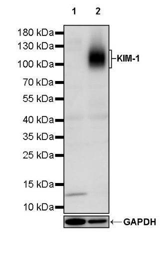
WB result of KIM-1 Rabbit mAb
Primary antibody: KIM-1 Rabbit mAb at 1/1000 dilution
Lane 1: HeLa whole cell lysate 20 µg
Lane 2: A549 whole cell lysate 20 µg
Negative control: HeLa whole cell lysate
Secondary antibody: Goat Anti-rabbit IgG, (H+L), HRP conjugated at 1/10000 dilution
Predicted MW: 39 kDa
Observed MW: 100~120 kDa
Flow cytometric analysis of HeLa (Human cervix adenocarcinoma epithelial cell, left) / A549 (Human lung carcinoma epithelial cell, right) cells labelling KIM-1 antibody at 1/50 dilution (1 μg)/ (Red) compared with a Rabbit monoclonal IgG (Black) isotype control and an unlabelled control (cells without incubation with primary antibody and secondary antibody) (Blue). Goat Anti - Rabbit IgG Alexa Fluor® 488 was used as the secondary antibody.
Negative control: HeLa
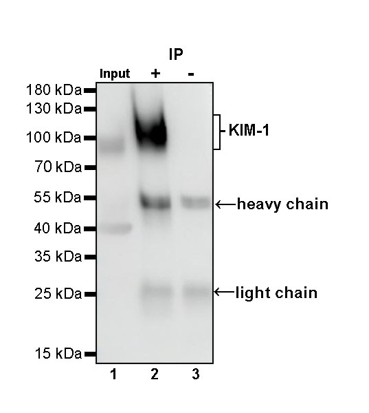
KIM-1 Rabbit mAb at 1/50 dilution (1 µg) immunoprecipitating KIM-1 in 0.4 mg A549 whole cell lysate.
Western blot was performed on the immunoprecipitate using KIM-1 Rabbit mAb at 1/1000 dilution.
Secondary antibody (HRP) for IP was used at 1/1000 dilution.
Lane 1: A549 whole cell lysate 20 µg (Input)
Lane 2: KIM-1 Rabbit mAb IP in A549 whole cell lysate
Lane 3: Rabbit monoclonal IgG IP in A549 whole cell lysate
Predicted MW: 39 kDa
Observed MW: 100~120 kDa
This blot was developed with high sensitivity substrate
IHC shows positive staining in paraffin-embedded human kidney. Anti-KIM-1 antibody was used at 1/500 dilution, followed by a HRP Polymer for Mouse & Rabbit IgG (ready to use). Counterstained with hematoxylin. Heat mediated antigen retrieval with Tris/EDTA buffer pH9.0 was performed before commencing with IHC staining protocol.
IHC shows positive staining in paraffin-embedded human hepatocellular carcinoma. Anti-KIM-1 antibody was used at 1/500 dilution, followed by a HRP Polymer for Mouse & Rabbit IgG (ready to use). Counterstained with hematoxylin. Heat mediated antigen retrieval with Tris/EDTA buffer pH9.0 was performed before commencing with IHC staining protocol.
ICC shows positive staining in A549 cells. Anti-KIM-1 antibody was used at 1/500 dilution (Green) and incubated overnight at 4°C. Goat polyclonal Antibody to Rabbit IgG - H&L (Alexa Fluor® 488) was used as secondary antibody at 1/1000 dilution. The cells were fixed with 4% PFA and permeabilized with 0.1% PBS-Triton X-100. Nuclei were counterstained with DAPI (Blue). Counterstain with tubulin (Red).
Negative control: ICC shows negative staining in HeLa cells. Anti-KIM-1 antibody was used at 1/500 dilution and incubated overnight at 4°C. Goat polyclonal Antibody to Rabbit IgG - H&L (Alexa Fluor® 488) was used as secondary antibody at 1/1000 dilution. The cells were fixed with 4% PFA and permeabilized with 0.1% PBS-Triton X-100. Nuclei were counterstained with DAPI (Blue). Counterstain with tubulin (Red).
 您现在的位置: