
12 months from date of receipt / reconstitution, -20 °C as supplied
| 应用 | 稀释度 |
|---|---|
| WB | 1:1000 |
| IP | 1:50 |
Akt, also known as Protein kinase B (PKB), is the collective name of a set of three serine/threonine-specific protein kinases that play key roles in multiple cellular processes such as glucose metabolism, apoptosis, cell proliferation, transcription, and cell migration. Once correctly positioned at the membrane via binding of PIP3, Akt can then be phosphorylated by its activating kinases, phosphoinositide-dependent kinase-1 (PDPK1 at threonine 308 in Akt1 and threonine 309 in Akt2) and the mammalian target of rapamycin complex 2 (mTORC2 at serine 473 (Akt1) and 474 (Akt2)). Activated Akt isoforms can then go on to activate or deactivate their myriad substrates (e.g. mTOR) via their kinase activity. The Akt kinases regulate cellular survival and metabolism by binding and regulating many downstream effectors, e.g. Nuclear Factor-κB, Bcl-2 family proteins, master lysosomal regulator TFEB and murine double minute 2 (MDM2).
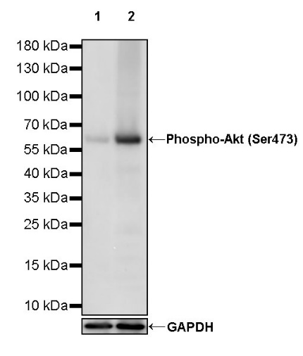
WB result of Phospho-Akt (Ser473) Rabbit mAb
Primary antibody: Phospho-Akt (Ser473) Rabbit mAb at 1/1000 dilution
Lane 1: untreated Jurkat whole cell lysate 20 µg
Lane 2: Jurkat treated with Calyculin A (100 nM, 30 mins) whole cell lysate 20 µg
Secondary antibody: Goat Anti-rabbit IgG, (H+L), HRP conjugated at 1/10000 dilution
Predicted MW: 56 kDa
Observed MW: 60 kDa
WB result of Phospho-Akt (Ser473) Rabbit mAb
Primary antibody: Phospho-Akt (Ser473) Rabbit mAb at 1/1000 dilution
Lane 1: untreated NIH/3T3 whole cell lysate 20 µg
Lane 2: NIH/3T3 treated with Calyculin A (100 nM, 30 mins) whole cell lysate 20 µg
Secondary antibody: Goat Anti-rabbit IgG, (H+L), HRP conjugated at 1/10000 dilution
Predicted MW: 56 kDa
Observed MW: 60 kDa
WB result of Phospho-Akt (Ser473) Rabbit mAb
Primary antibody: Phospho-Akt (Ser473) Rabbit mAb at 1/1000 dilution
Lane 1: untreated C6 whole cell lysate 20 µg
Lane 2: C6 treated with Calyculin A (100 nM, 30 mins) whole cell lysate 20 µg
Secondary antibody: Goat Anti-rabbit IgG, (H+L), HRP conjugated at 1/10000 dilution
Predicted MW: 56 kDa
Observed MW: 60 kDa
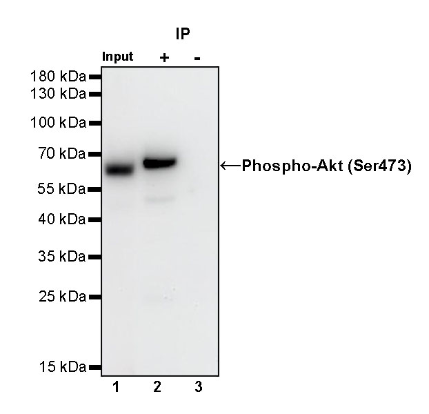
Phospho-Akt (Ser473) Rabbit mAb at 1/50 dilution (1 µg) immunoprecipitating Phospho-Akt (Ser473) in 0.4 mg Jurkat treated with Calyculin A (100 nM, 30 mins) whole cell lysate.
Western blot was performed on the immunoprecipitate using Phospho-Akt (Ser473) Rabbit mAb at 1/1000 dilution.
Secondary antibody (HRP) for IP was used at 1/1000 dilution.
Lane 1: Jurkat treated with Calyculin A (100 nM, 30 mins) whole cell lysate 20 µg (Input)
Lane 2: Phospho-Akt (Ser473) Rabbit mAb IP in Jurkat treated with Calyculin A (100 nM, 30 mins) whole cell lysate
Lane 3: Rabbit monoclonal IgG IP in Jurkat treated with Calyculin A (100 nM, 30 mins) whole cell lysate
Predicted MW: 56 kDa
Observed MW: 60 kDa
This blot was developed with high sensitivity substrate
Dot blot result of Phospho-Akt (Ser473) Rabbit mAb
Lane 1: Phospho-Akt (Ser473) peptide
Lane 2: Akt unmodified peptide
Primary antibody: Phospho-Akt (Ser473) Rabbit mAb at 1/1000 dilution
Secondary antibody: Goat Anti-Rabbit IgG, (H+L), HRP conjugated at 1/10000 dilution
 您现在的位置: