
12 months from date of receipt / reconstitution, -20 °C as supplied
| 应用 | 稀释度 |
|---|---|
| WB | 1:1000 |
| IP | 1:50 |
| IHC-P | 1:500 |
| ICC | 1:500 |
| ICFCM | 1:500 |
Pyruvate kinase is the enzyme involved in the last step of glycolysis. It catalyzes the transfer of a phosphate group from phosphoenolpyruvate (PEP) to adenosine diphosphate (ADP), yielding one molecule of pyruvate and one molecule of ATP. In contrast to mitochondrial respiration, energy regeneration by pyruvate kinase is independent from oxygen supply and allows survival of the organs under hypoxic conditions often found in solid tumors. Pyruvate kinase is present in four distinct, tissue-specific isozymes in animals, each consisting of particular kinetic properties necessary to accommodate the variations in metabolic requirements of diverse tissues.
WB result of PKM Rabbit mAb
Primary antibody: PKM Rabbit mAb at 1/1000 dilution
Lane 1: HeLa whole cell lysate 20 µg
Lane 2: Jurkat whole cell lysate 20 µg
Lane 3: A549 whole cell lysate 20 µg
Secondary antibody: Goat Anti-rabbit IgG, (H+L), HRP conjugated at 1/10000 dilution
Predicted MW: 58 kDa
Observed MW: 60 kDa
(This blot was developed with high sensitivity substrate)
WB result of PKM Rabbit mAb
Primary antibody: PKM Rabbit mAb at 1/1000 dilution
Lane 1: NIH/3T3 whole cell lysate 20 µg
Lane 2: mouse lung lysate 20 µg
Lane 3: mouse skeletal muscle lysate 20 µg
Lane 4: mouse brain lysate 20 µg
Lane 5: mouse spleen lysate 20 µg
Lane 6: mouse testis lysate 20 µg
Secondary antibody: Goat Anti-rabbit IgG, (H+L), HRP conjugated at 1/10000 dilution
Predicted MW: 58 kDa
Observed MW: 60 kDa
WB result of PKM Rabbit mAb
Primary antibody: PKM Rabbit mAb at 1/1000 dilution
Lane 1: rat skeletal muscle lysate 20 µg
Lane 2: rat brain lysate 20 µg
Lane 3: rat testis lysate 20 µg
Lane 4: rat spleen lysate 20 µg
Secondary antibody: Goat Anti-rabbit IgG, (H+L), HRP conjugated at 1/10000 dilution
Predicted MW: 58 kDa
Observed MW: 60 kDa
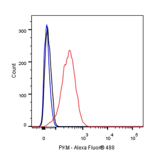
Flow cytometric analysis of 4% PFA fixed 90% methanol permeabilized HeLa (Human cervix adenocarcinoma epithelial cell) cells labelling α-tubulin antibody at 1/500 dilution (0.1 μg) / (red) compared with a Rabbit monoclonal IgG (Black) isotype control and an unlabelled control (cells without incubation with primary antibody and secondary antibody) (Blue). Goat Anti - Rabbit IgG Alexa Fluor® 488 was used as the secondary antibody.
PKM Rabbit mAb at 1/50 dilution (1 µg) immunoprecipitating PKM in 0.4 mg HeLa whole cell lysate.
Western blot was performed on the immunoprecipitate using PKM Rabbit mAb at 1/1000 dilution.
Secondary antibody (HRP) for IP was used at 1/400 dilution.
Lane 1: HeLa whole cell lysate 10 µg (Input)
Lane 2: PKM Rabbit mAb IP in HeLa whole cell lysate
Lane 3: Rabbit monoclonal IgG IP in HeLa whole cell lysate
Predicted MW: 58 kDa
Observed MW: 60 kDa
IHC shows positive staining in paraffin-embedded human kidney. Anti-PKM antibody was used at 1/500 dilution, followed by a HRP Polymer for Mouse & Rabbit IgG (ready to use). Counterstained with hematoxylin. Heat mediated antigen retrieval with Tris/EDTA buffer pH9.0 was performed before commencing with IHC staining protocol.
IHC shows positive staining in paraffin-embedded human cervical squamous cell carcinoma. Anti-PKM antibody was used at 1/500 dilution, followed by a HRP Polymer for Mouse & Rabbit IgG (ready to use). Counterstained with hematoxylin. Heat mediated antigen retrieval with Tris/EDTA buffer pH9.0 was performed before commencing with IHC staining protocol.
IHC shows positive staining in paraffin-embedded human endometrial carcinoma. Anti-PKM antibody was used at 1/500 dilution, followed by a HRP Polymer for Mouse & Rabbit IgG (ready to use). Counterstained with hematoxylin. Heat mediated antigen retrieval with Tris/EDTA buffer pH9.0 was performed before commencing with IHC staining protocol.
IHC shows positive staining in paraffin-embedded mouse cerebral cortex. Anti-PKM antibody was used at 1/500 dilution, followed by a HRP Polymer for Mouse & Rabbit IgG (ready to use). Counterstained with hematoxylin. Heat mediated antigen retrieval with Tris/EDTA buffer pH9.0 was performed before commencing with IHC staining protocol.
IHC shows positive staining in paraffin-embedded rat cerebral cortex. Anti-PKM antibody was used at 1/500 dilution, followed by a HRP Polymer for Mouse & Rabbit IgG (ready to use). Counterstained with hematoxylin. Heat mediated antigen retrieval with Tris/EDTA buffer pH9.0 was performed before commencing with IHC staining protocol.
ICC shows positive staining in HeLa cells. Anti-PKM antibody was used at 1/500 dilution (Green) and incubated overnight at 4°C. Goat polyclonal Antibody to Rabbit IgG - H&L (Alexa Fluor® 488) was used as secondary antibody at 1/1000 dilution. The cells were fixed with 4% PFA and permeabilized with 0.1% PBS-Triton X-100. Nuclei were counterstained with DAPI (Blue). Counterstain with tubulin (Red).
 您现在的位置: