
PBS, 40% Glycerol, 0.05% BSA, 0.03% Proclin 300
12 months from date of receipt / reconstitution, -20 °C as supplied
| 应用 | 稀释度 |
|---|---|
| WB | 1:1000 |
| IP | 1:50 |
| ICC | 1:100-1:500 |
| ICFCM | 1:500 |
SIRT6 is member of the mammalian sirtuin family of proteins, which are homologs to the yeast Sir2 protein. Sirt6 is mainly known as a deacetylase of histones H3 and H4, an activity by which it changes chromatin density and regulates gene expression. The enzymatic activity of Sirt6, as well as of the other members of the sirtuins family, is dependent upon the binding of the cofactor nicotinamide adenine dinucleotide (NAD+). SIRT6 is a chromatin-associated protein that is required for normal base excision repair and double-strand break repair of DNA damage in mammalian cells. SIRT6 promotes the repair of DNA double-strand breaks by the process of non-homologous end joining and homologous recombination. SIRT6 stabilizes the repair protein DNA-PKcs (DNA-dependent protein kinase catalytic subunit) at chromatin sites of damage.
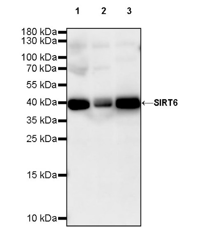
WB result of SIRT6 Rabbit mAb
Primary antibody: SIRT6 Rabbit mAb at 1/1000 dilution
Lane 1: HCT 116 whole cell lysate 20 µg
Lane 2: HeLa whole cell lysate 20 µg
Lane 3: Jurkat whole cell lysate 20 µg
Secondary antibody: Goat Anti-rabbit IgG, (H+L), HRP conjugated at 1/10000 dilution
Predicted MW: 39 kDa
Observed MW: 40 kDa
(This blot was developed with high sensitivity substrate)
Flow cytometric analysis of 4% PFA fixed 90% methanol permeabilized HeLa (Human cervix adenocarcinoma epithelial cell) cells labelling SIRT6 antibody at 1/500 dilution (0.1 μg) / (Red) compared with a Rabbit monoclonal IgG (Black) isotype control and an unlabelled control (cells without incubation with primary antibody and secondary antibody) (Blue). Goat Anti - Rabbit IgG Alexa Fluor® 488 was used as the secondary antibody.
SIRT6 Rabbit mAb at 1/50 dilution (1 µg) immunoprecipitating SIRT6 in 0.4 mg HCT 116 whole cell lysate.
Western blot was performed on the immunoprecipitate using SIRT6 Rabbit mAb at 1/1000 dilution.
Secondary antibody (HRP) for IP was used at 1/400 dilution.
Lane 1: HCT 116 whole cell lysate 20 µg (Input)
Lane 2: SIRT6 Rabbit mAb IP in HCT 116 whole cell lysate
Lane 3: Rabbit monoclonal IgG IP in HCT 116 whole cell lysate
Predicted MW: 39 kDa
Observed MW: 39 kDa
ICC shows positive staining in HeLa cells. Anti-SIRT6 antibody was used at 1/500 dilution (Green) and incubated overnight at 4°C. Goat polyclonal Antibody to Rabbit IgG - H&L (Alexa Fluor® 488) was used as secondary antibody at 1/1000 dilution. The cells were fixed with 4% PFA and permeabilized with 0.1% PBS-Triton X-100. Nuclei were counterstained with DAPI (Blue). Counterstain with tubulin (Red).
ICC shows positive staining in HCT-116 cells. Anti-SIRT6 antibody was used at 1/100 dilution (Green) and incubated overnight at 4°C. Goat polyclonal Antibody to Rabbit IgG - H&L (Alexa Fluor® 488) was used as secondary antibody at 1/1000 dilution. The cells were fixed with 100% ice-cold methanol and permeabilized with 0.1% PBS-Triton X-100. Nuclei were counterstained with DAPI (Blue). Counterstain with tubulin (Red).
 您现在的位置: