
12 months from date of receipt / reconstitution, -20 °C as supplied
| 应用 | 稀释度 |
|---|---|
| WB | 1:1000 |
| IP | 1:50 |
| IHC-P | 1:250 |
| ICC | 1:500 |
| ICFCM | 1:500 |
Thymidylate synthase (TS) is an enzyme that catalyzes the conversion of deoxyuridine monophosphate (dUMP) to deoxythymidine monophosphate (dTMP). TS has become an important target for cancer treatment by means of chemotherapy. The sensitivity of TS to succumb to TS inhibitors is a key part to its success as treatment for colorectal, pancreatic, ovarian, gastric, and breast cancers.
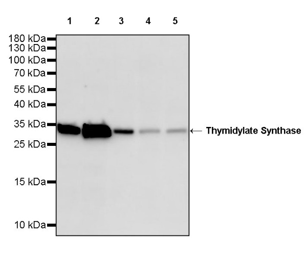
WB result of Thymidylate Synthase Rabbit mAb
Primary antibody: Thymidylate Synthase Rabbit mAb at 1/1000 dilution
Lane 1: HeLa whole cell lysate 20 µg
Lane 2: Jurkat whole cell lysate 20 µg
Lane 3: SK-BR-3 whole cell lysate 20 µg
Lane 4: Hep G2 whole cell lysate 20 µg
Lane 5: MCF7 whole cell lysate 20 µg
Secondary antibody: Goat Anti-rabbit IgG, (H+L), HRP conjugated at 1/10000 dilution
Predicted MW: 35 kDa
Observed MW: 35 kDa
WB result of Thymidylate Synthase Rabbit mAb
Primary antibody: Thymidylate Synthase Rabbit mAb at 1/1000 dilution
Lane 1: NIH/3T3 whole cell lysate 20 µg
Lane 2: RAW 264.7 whole cell lysate 20 µg
Secondary antibody: Goat Anti-rabbit IgG, (H+L), HRP conjugated at 1/10000 dilution
Predicted MW: 35 kDa
Observed MW: 35 kDa
WB result of Thymidylate Synthase Rabbit mAb
Primary antibody: Thymidylate Synthase Rabbit mAb at 1/1000 dilution
Lane 1: C6 whole cell lysate 20 µg
Secondary antibody: Goat Anti-rabbit IgG, (H+L), HRP conjugated at 1/10000 dilution
Predicted MW: 35 kDa
Observed MW: 35 kDa
Flow cytometric analysis of 4% PFA fixed 90% methanol permeabilized Jurkat (Human T cell leukemia T lymphocyte) cells labelling Thymidylate Synthase antibody at 1/500 dilution (0.1 μg) / (Red) compared with a Rabbit monoclonal IgG (Black) isotype control and an unlabelled control (cells without incubation with primary antibody and secondary antibody) (Blue). Goat Anti - Rabbit IgG Alexa Fluor® 488 was used as the secondary antibody.
Thymidylate Synthase Rabbit mAb at 1/50 dilution (1 µg) immunoprecipitating Thymidylate Synthase in 0.4 mg HeLa whole cell lysate.
Western blot was performed on the immunoprecipitate using Thymidylate Synthase Rabbit mAb at 1/1000 dilution.
Secondary antibody (HRP) for IP was used at 1/400 dilution.
Lane 1: HeLa whole cell lysate 20 µg (Input)
Lane 2: Thymidylate Synthase Rabbit mAb IP in HeLa whole cell lysate
Lane 3: Rabbit monoclonal IgG IP in HeLa whole cell lysate
Predicted MW: 36 kDa
Observed MW: 34 kDa
IHC shows positive staining in paraffin-embedded human tonsil. Anti-Thymidylate Synthase antibody was used at 1/250 dilution, followed by a HRP Polymer for Mouse & Rabbit IgG (ready to use). Counterstained with hematoxylin. Heat mediated antigen retrieval with Tris/EDTA buffer pH9.0 was performed before commencing with IHC staining protocol.
IHC shows positive staining in paraffin-embedded human spleen. Anti-Thymidylate Synthase antibody was used at 1/250 dilution, followed by a HRP Polymer for Mouse & Rabbit IgG (ready to use). Counterstained with hematoxylin. Heat mediated antigen retrieval with Tris/EDTA buffer pH9.0 was performed before commencing with IHC staining protocol.
ICC shows positive staining in Jurkat cells. Anti- Thymidylate Synthase antibody was used at 1/500 dilution (Green) and incubated overnight at 4°C. Goat polyclonal Antibody to Rabbit IgG - H&L (Alexa Fluor® 488) was used as secondary antibody at 1/1000 dilution. The cells were fixed with 100% ice-cold methanol and permeabilized with 0.1% PBS-Triton X-100. Nuclei were counterstained with DAPI (Blue). Counterstain with tubulin (Red).
 您现在的位置: