
12 months from date of receipt / reconstitution, -20 °C as supplied
| 应用 | 稀释度 |
|---|---|
| WB | 1:1000 |
| IHC-P | 1:50-1:400 |
| ICC | 1:500 |
| ICFCM | 1:500 |
Chitinase-3 like-protein-1 (CHI3L1), one of the CLPs, also has been named YKL-40 in humans and breast regression protein 39 (BRP-39) in mice, is common in both prokaryotes and eukaryotes. Following its initial discovery in the culture supernatant of the osteosarcoma cell line MG63, it was subsequently detected in human chondrocytes, synoviocytes, and vascular smooth muscle cells. In fact, CHI3L1 is produced by a multitude of cells, including macrophages, neutrophils, fibroblast-like cells, hepatic stellate cells, endothelial cells, and cancer cells. CHI3L1 is overexpressed in many human cancer types and animal tumor models, for instance, oligodendroglia, glioblastoma, osteosarcoma, sarcoma, colon, and gastric cancers (GCs). Elevated serum levels of CHI3L1 have been found to be associated with poor prognosis and shorter survival in patients with metastatic cancer. Consequently, CHI3L1 has been increasingly proposed as a sensitive biomarker and an attractive therapeutic target for several certain types of cancers.
WB result of CHI3L1 Rabbit mAb
Primary antibody: CHI3L1 Rabbit mAb at 1/1000 dilution
Lane 1: 293T whole cell lysate 20 µg
Lane 2: THP-1 whole cell lysate 20 µg
Negative control: 293T whole cell lysate
Secondary antibody: Goat Anti-Rabbit IgG, (H+L), HRP conjugated at 1/10000 dilution
Predicted MW: 43 kDa
Observed MW: 40, 37 kDa
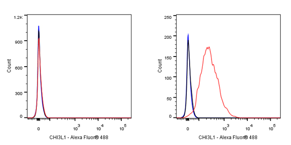
Flow cytometric analysis of 4% PFA fixed 90% methanol permeabilized 293T (Human embryonic kidney epithelial cell, left) / THP-1 (Human monocytic leukemia monocyte, Right) labelling CHI3L1 antibody at 1/500 dilution (0.1 μg) / (Red) compared with a Rabbit monoclonal IgG (Black) isotype control and an unlabelled control (cells without incubation with primary antibody and secondary antibody) (Blue). Goat Anti - Rabbit IgG Alexa Fluor® 488 was used as the secondary antibody.
Negative control: 293T
IHC shows positive staining in paraffin-embedded human kidney. Anti-CHI3L1 antibody was used at 1/400 dilution, followed by a HRP Polymer for Mouse & Rabbit IgG (ready to use). Counterstained with hematoxylin. Heat mediated antigen retrieval with Tris/EDTA buffer pH9.0 was performed before commencing with IHC staining protocol.
IHC shows positive staining in paraffin-embedded human spleen. Anti-CHI3L1 antibody was used at 1/50 dilution, followed by a HRP Polymer for Mouse & Rabbit IgG (ready to use). Counterstained with hematoxylin. Heat mediated antigen retrieval with Tris/EDTA buffer pH9.0 was performed before commencing with IHC staining protocol.
IHC shows positive staining in paraffin-embedded human tonsil. Anti-CHI3L1 antibody was used at 1/100 dilution, followed by a HRP Polymer for Mouse & Rabbit IgG (ready to use). Counterstained with hematoxylin. Heat mediated antigen retrieval with Tris/EDTA buffer pH9.0 was performed before commencing with IHC staining protocol.
IHC shows positive staining in paraffin-embedded human colon cancer. Anti-CHI3L1 antibody was used at 1/100 dilution, followed by a HRP Polymer for Mouse & Rabbit IgG (ready to use). Counterstained with hematoxylin. Heat mediated antigen retrieval with Tris/EDTA buffer pH9.0 was performed before commencing with IHC staining protocol.
ICC shows positive staining in THP-1 cells. Anti-CHI3L1 antibody was used at 1/500 dilution (Green) and incubated overnight at 4°C. Goat polyclonal Antibody to Rabbit IgG - H&L (Alexa Fluor® 488) was used as secondary antibody at 1/1000 dilution. The cells were fixed with 100% ice-cold methanol and permeabilized with 0.1% PBS-Triton X-100. Nuclei were counterstained with DAPI (Blue).
Negative control: ICC shows negative staining in 293T cells. Anti-CHI3L1 antibody was used at 1/500 dilution and incubated overnight at 4°C. Goat polyclonal Antibody to Rabbit IgG - H&L (Alexa Fluor® 488) was used as secondary antibody at 1/1000 dilution. The cells were fixed with 100% ice-cold methanol and permeabilized with 0.1% PBS-Triton X-100. Nuclei were counterstained with DAPI (Blue).
 您现在的位置: