
12 months from date of receipt / reconstitution, -20°C as supplied
| 应用 | 稀释度 |
|---|---|
| WB | 1:1000 |
| ICC | 1:500 |
| ICFCM | 1:500 |
CEBP beta is a bZIP transcription factor that can bind as a homodimer to certain DNA regulatory regions. It can also form heterodimers with the related proteins CEBP-alpha, CEBP-delta, and CEBP-gamma. It is important in the regulation of genes involved in immune and inflammatory responses and has been shown to bind to the IL-1 response element in the IL-6 gene, as well as to regulatory regions of several acute-phase and cytokine genes. In addition, CEBP beta can bind the promoter and upstream element and stimulate the expression of the collagen type I gene.
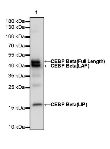
WB result of CEBP Beta (C-terminal) Rabbit mAb
Primary antibody: CEBP Beta (C-terminal) Rabbit mAb at 1/1000 dilution
Lane 1: HeLa whole cell lysate 20 µg
Secondary antibody: Goat Anti-Rabbit IgG, (H+L), HRP conjugated at 1/10000 dilution
Predicted MW: 38 kDa
Observed MW: 20, 40, 45 kDa
(This blot was developed with high sensitivity substrate)
WB result of CEBP Beta (C-terminal) Rabbit mAb
Primary antibody: CEBP Beta (C-terminal) Rabbit mAb at 1/1000 dilution
Lane 1: NIH/3T3 whole cell lysate 20 µg
Secondary antibody: Goat Anti-Rabbit IgG, (H+L), HRP conjugated at 1/10000 dilution
Predicted MW: 38 kDa
Observed MW: 20, 35, 38 kDa
(This blot was developed with high sensitivity substrate)
WB result of CEBP Beta (C-terminal) Rabbit mAb
Primary antibody: CEBP Beta (C-terminal) Rabbit mAb at 1/1000 dilution
Lane 1: PC-12 whole cell lysate 20 µg
Secondary antibody: Goat Anti-Rabbit IgG, (H+L), HRP conjugated at 1/10000 dilution
Predicted MW: 38 kDa
Observed MW: 20, 35, 38 kDa
(This blot was developed with high sensitivity substrate)
Flow cytometric analysis of 4% PFA fixed 90% methanol permeabilized HeLa (Human cervix adenocarcinoma epithelial cell) cells labelling CEBP Beta (C-terminal) antibody at 1/500 dilution (0.1 μg)/ (Red) compared with a Rabbit monoclonal IgG (Black) isotype control and an unlabelled control (cells without incubation with primary antibody and secondary antibody) (Blue). Goat Anti - Rabbit IgG Alexa Fluor® 488 was used as the secondary antibody.
Flow cytometric analysis of 4% PFA fixed 90% methanol permeabilized NIH/3T3 (Mouse embryonic fibroblast) cells labelling CEBP Beta (C-terminal) antibody at 1/500 dilution (0.1 μg)/ (Red) compared with a Rabbit monoclonal IgG (Black) isotype control and an unlabelled control (cells without incubation with primary antibody and secondary antibody) (Blue). Goat Anti - Rabbit IgG Alexa Fluor® 488 was used as the secondary antibody.
ICC shows positive staining in HeLa cells. Anti- CEBP Beta (C-terminal) antibody was used at 1/500 dilution (Green) and incubated overnight at 4°C. Goat polyclonal Antibody to Rabbit IgG - H&L (Alexa Fluor® 488) was used as secondary antibody at 1/1000 dilution. The cells were fixed with 4% PFA and permeabilized with 0.1% PBS-Triton X-100. Nuclei were counterstained with DAPI (Blue). Counterstain with tubulin (Red).
ICC shows positive staining in NIH/3T3 cells. Anti-CEBP Beta (C-terminal) antibody was used at 1/500 dilution (Green) and incubated overnight at 4°C. Goat polyclonal Antibody to Rabbit IgG - H&L (Alexa Fluor® 488) was used as secondary antibody at 1/1000 dilution. The cells were fixed with 4% PFA and permeabilized with 0.1% PBS-Triton X-100. Nuclei were counterstained with DAPI (Blue). Counterstain with tubulin (Red).
 您现在的位置: