
PBS, 40% Glycerol, 0.05% BSA, 0.03% Proclin 300
12 months from date of receipt / reconstitution, -20 °C as supplied
| 应用 | 稀释度 |
|---|---|
| WB | 1:1000 |
| ICC | 1:100 |
| FCM | 1:2000 |
Programmed cell death protein 1, also known as PD-1 and CD279 (cluster of differentiation 279), is a protein on the surface of T and B cells that has a role in regulating the immune system's response to the cells of the human body by down-regulating the immune system and promoting self-tolerance by suppressing T cell inflammatory activity. This prevents autoimmune diseases, but it can also prevent the immune system from killing cancer cells. PD-1 is an immune checkpoint and guards against autoimmunity through two mechanisms. First, it promotes apoptosis (programmed cell death) of antigen-specific T-cells in lymph nodes. Second, it reduces apoptosis in regulatory T cells (anti-inflammatory, suppressive T cells).
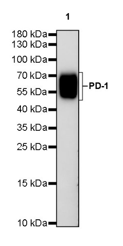
WB result of PD-1 (mouse) Rat mAb
Primary antibody: PD-1 (mouse) Rat mAb at 1/1000 dilution
Lane 1: recombinant PD-1 Fc Chimera 1 µg
Secondary antibody: Goat Anti-Rat IgG, (H+L), HRP conjugated at 1/10000 dilution
Predicted MW: 31 kDa
Observed MW: 50~70 kDa
(This blot was developed with high sensitivity substrate)
Flow cytometric analysis of NIH/3T3 (Mouse embryonic fibroblast, left) / EL4.IL-2 (Mouse lymphoma T lymphocyte, Right) labelling PD-1 (mouse) antibody at 1/2000 dilution (0.1 μg) / (Red) compared with a Rat monoclonal IgG (Black) isotype control and an unlabelled control (cells without incubation with primary antibody and secondary antibody) (Blue). Goat Anti – Rat IgG Alexa Fluor® 488 was used as the secondary antibody.
Negative control: NIH/3T3
ICC shows positive staining in EL4.IL2 cells. Anti- PD-1 antibody was used at 1/100 dilution (Green) and incubated overnight at 4°C. Goat polyclonal Antibody to Rat IgG - H&L (Alexa Fluor® 488) was used as secondary antibody at 1/1000 dilution. The cells were fixed with 4% PFA and permeabilized with 0.1% PBS-Triton X-100. Nuclei were counterstained with DAPI (Blue).
Negative control: ICC shows negative staining in NIH/3T3 cells. Anti- PD-1 antibody was used at 1/100 dilution and incubated overnight at 4°C. Goat polyclonal Antibody to Rat IgG - H&L (Alexa Fluor® 488) was used as secondary antibody at 1/1000 dilution. The cells were fixed with 4% PFA and permeabilized with 0.1% PBS-Triton X-100. Nuclei were counterstained with DAPI (Blue).
 您现在的位置: