
12 months from date of receipt / reconstitution, -20 °C as supplied
| 应用 | 稀释度 |
|---|---|
| WB | 1:1000 |
| IP | 1:50 |
| ICC | 1:100 |
| ICFCM | 1:500 |
The TATA-binding protein (TBP) is a general transcription factor that binds specifically to a DNA sequence called the TATA box. This DNA sequence is found about 30 base pairs upstream of the transcription start site in some eukaryotic gene promoters. TBP is a subunit of the eukaryotic general transcription factor TFIID. TFIID is the first protein to bind to DNA during the formation of the transcription preinitiation complex of RNA polymerase II (RNA Pol II). As one of the few proteins in the preinitiation complex that binds DNA in a sequence-specific manner, it helps position RNA polymerase II over the transcription start site of the gene. TBP is also involved in DNA melting (double strand separation) by bending the DNA by 80° (the AT-rich sequence to which it binds facilitates easy melting). The TBP is an unusual protein in that it binds the minor groove using a β sheet.
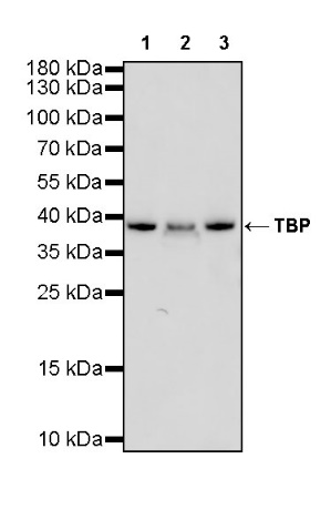
WB result of TBP Rabbit mAb
Primary antibody: TBP Rabbit mAb at 1/1000 dilution
Lane 1: HeLa whole cell lysate 20 µg
Lane 2: 293T whole cell lysate 20 µg
Lane 3: Hep G2 whole cell lysate 20 µg
Secondary antibody: Goat Anti-Rabbit IgG, (H+L), HRP conjugated at 1/10000 dilution
Predicted MW: 38 kDa
Observed MW: 38 kDa
WB result of TBP Rabbit mAb
Primary antibody: TBP Rabbit mAb at 1/1000 dilution
Lane 1: PC-12 whole cell lysate 20 µg
Secondary antibody: Goat Anti-Rabbit IgG, (H+L), HRP conjugated at 1/10000 dilution
Predicted MW: 38 kDa
Observed MW: 38 kDa
Flow cytometric analysis of 4% PFA fixed 90% methanol permeabilized HeLa (Human cervix adenocarcinoma epithelial cell) cells labelling TBP antibody at 1/500 dilution (0.1 μg)/ (Red) compared with a Rabbit monoclonal IgG (Black) isotype control and an unlabelled control (cells without incubation with primary antibody and secondary antibody) (Blue). Goat Anti - Rabbit IgG Alexa Fluor® 488 was used as the secondary antibody.
TBP Rabbit mAb at 1/50 dilution (1 µg) immunoprecipitating TBP in 0.4 mg HeLa whole cell lysate.
Western blot was performed on the immunoprecipitate using TBP Rabbit mAb at 1/1000 dilution.
Secondary antibody (HRP) for IP was used at 1/400 dilution.
Lane 1: HeLa whole cell lysate 20 µg (Input)
Lane 2: TBP Rabbit mAb IP in HeLa whole cell lysate
Lane 3: Rabbit monoclonal IgG IP in HeLa whole cell lysate
Predicted MW: 38 kDa
Observed MW: 38 kDa
ICC shows positive staining in HeLa cells. Anti-TBP antibody was used at 1/100 dilution (Green) and incubated overnight at 4°C. Goat polyclonal Antibody to Rabbit IgG - H&L (Alexa Fluor® 488) was used as secondary antibody at 1/1000 dilution. The cells were fixed with 4% PFA and permeabilized with 0.1% PBS-Triton X-100. Nuclei were counterstained with DAPI (Blue). Counterstain with tubulin (Red).
 您现在的位置: