
12 months from date of receipt / reconstitution, -20 °C as supplied
| 应用 | 稀释度 |
|---|---|
| WB | 1:1000~1:5000 |
| IHC-P | 1:500 |
| ICC | 1:1000 |
| ICFCM | 1:2000 |
Tubulin is the major constituent of microtubules, a cylinder consisting of laterally associated linear protofilaments composed of alpha- and beta-tubulin heterodimers. Tubulin α- and β-subunits have molecular weights of ~ 50 kDa and are 36%–42% identical and 63% homologous. Both tubulin subunits bind guanine nucleotides. The binding to α-tubulin at the N-site is nonexchangeable, while the binding to β-tubulin at the E-site is exchangeable. Nucleotide in microtubules does not exchange with the solution, except for terminal subunits at microtubule ends.
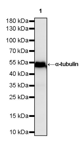
WB result of α-tubulin Mouse mAb
Primary antibody: α-tubulin Mouse mAb at 1/1000 dilution
Lane 1: HeLa whole cell lysate 20 µg
Secondary antibody: Goat Anti-Mouse IgG, (H+L), HRP conjugated at 1/10000 dilution
Predicted MW: 55 kDa
Observed MW: 52 kDa
WB result of α-tubulin Mouse mAb
Primary antibody: α-tubulin Mouse mAb at 1/1000 dilution
Lane 1: NIH/3T3 whole cell lysate 20 µg
Secondary antibody: Goat Anti-Mouse IgG, (H+L), HRP conjugated at 1/10000 dilution
Predicted MW: 55 kDa
Observed MW: 52 kDa
WB result of α-tubulin Mouse mAb
Primary antibody: α-tubulin Mouse mAb at 1/1000 dilution
Lane 1: C6 whole cell lysate 20 µg
Secondary antibody: Goat Anti-Mouse IgG, (H+L), HRP conjugated at 1/10000 dilution
Predicted MW: 55 kDa
Observed MW: 52 kDa
WB result of α-tubulin Mouse mAb
Primary antibody: α-tubulin Mouse mAb at 1/5000 dilution
Lane 1: pig heart lysate 20 µg
Secondary antibody: Goat Anti-Mouse IgG, (H+L), HRP conjugated at 1/10000 dilution
Predicted MW: 55 kDa
Observed MW: 52 kDa
(This blot was developed with high sensitivity substrate)
WB result of α-tubulin Mouse mAb
Primary antibody : α-tubulin Mouse mAb at 1/1000 dilution
Lane 1 : Zebra fish lysate 20 µg
Secondary antibody: Goat Anti-Mouse IgG, (H+L), HRP conjugated at 1/10000 dilution
Predicted MW: 55 kDa
Observed MW: 52 kDa
Flow cytometric analysis of 4% PFA fixed 90% methanol permeabilized HeLa (Human cervix adenocarcinoma epithelial cell) cells labelling α-tubulin antibody at 1/2000 dilution (0.1 μg) / (Red) compared with a Mouse monoclonal IgG (Black) isotype control and an unlabelled control (cells without incubation with primary antibody and secondary antibody) (Blue). Goat Anti - Mouse IgG Alexa Fluor® 488 was used as the secondary antibody.
IHC shows positive staining in paraffin-embedded human kidney. Anti-α-tubulin antibody was used at 1/500 dilution, followed by a HRP Polymer for Mouse & Rabbit IgG (ready to use). Counterstained with hematoxylin. Heat mediated antigen retrieval with Tris/EDTA buffer pH9.0 was performed before commencing with IHC staining protocol.
IHC shows positive staining in paraffin-embedded human colon cancer. Anti-α-tubulin antibody was used at 1/500 dilution, followed by a HRP Polymer for Mouse & Rabbit IgG (ready to use). Counterstained with hematoxylin. Heat mediated antigen retrieval with Tris/EDTA buffer pH9.0 was performed before commencing with IHC staining protocol.
IHC shows positive staining in paraffin-embedded human endometrial cancer. Anti-α-tubulin antibody was used at 1/500 dilution, followed by a HRP Polymer for Mouse & Rabbit IgG (ready to use). Counterstained with hematoxylin. Heat mediated antigen retrieval with Tris/EDTA buffer pH9.0 was performed before commencing with IHC staining protocol.
IHC shows positive staining in paraffin-embedded mouse cerebral cortex. Anti-α-tubulin antibody was used at 1/500 dilution, followed by a HRP Polymer for Mouse & Rabbit IgG (ready to use). Counterstained with hematoxylin. Heat mediated antigen retrieval with Tris/EDTA buffer pH9.0 was performed before commencing with IHC staining protocol.
IHC shows positive staining in paraffin-embedded rat cerebral cortex. Anti-α-tubulin antibody was used at 1/500 dilution, followed by a HRP Polymer for Mouse & Rabbit IgG (ready to use). Counterstained with hematoxylin. Heat mediated antigen retrieval with Tris/EDTA buffer pH9.0 was performed before commencing with IHC staining protocol.
ICC shows positive staining in HeLa cells. Anti-α-tubulin antibody was used at 1/1000 dilution (Green) and incubated overnight at 4°C. Goat polyclonal Antibody to Mouse IgG - H&L (Alexa Fluor® 488) was used as secondary antibody at 1/1000 dilution. The cells were fixed with 100% ice-cold methanol and permeabilized with 0.1% PBS-Triton X-100. Nuclei were counterstained with DAPI (Blue).
 您现在的位置: