
PBS, 40% Glycerol, 0.05% BSA, 0.03% Proclin 300
12 months from date of receipt / reconstitution, -20 °C as supplied
| 应用 | 稀释度 |
|---|---|
| WB | 1:1000 |
| ICC | 1:500 |
| ICFCM | 1:500 |
FLAG-tag, or FLAG octapeptide, or FLAG epitope, is a peptide protein tag that can be added to a protein using recombinant DNA technology, having the sequence DYKDDDDK (where D=aspartic acid, Y=tyrosine, and K=lysine). It is one of the most specific tags and it is an artificial antigen to which specific, high affinity monoclonal antibodies have been developed and hence can be used for protein purification by affinity chromatography and also can be used for locating proteins within living cells. FLAG-tag has been used to separate recombinant, overexpressed protein from wild-type protein expressed by the host organism. FLAG-tag can also be used in the isolation of protein complexes with multiple subunits.
WB result of FLAG Tag Rabbit mAb
Primary antibody: FLAG Tag Rabbit mAb at 1/1000 dilution
Lane 1: 293-F whole cell lysate 20 µg
Lane 2: 293-F transfected with Flag tagged GFP whole cell Lysate 20 µg
Secondary antibody: Goat Anti-Rabbit IgG, (H+L), HRP conjugated at 1/10000 dilution
Predicted MW: 27 kDa
Observed MW: 32 kDa
Exposure time: 90 s
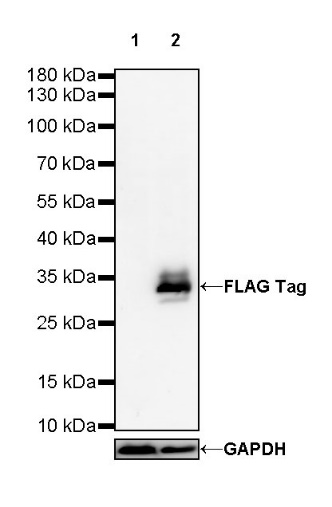
WB result of FLAG Tag Rabbit mAb
Primary antibody: FLAG Tag Rabbit mAb at 1/1000 dilution
Lane 1: Nicotiana benthamiana leaves (transfected with HA tagged GFP) lysate 20 µg
Lane 2: Nicotiana benthamiana leaves (transfected with Flag tagged GFP) lysate 20 µg
Secondary antibody: Goat Anti-Rabbit IgG, (H+L), HRP conjugated at 1/10000 dilution
Predicted MW: 27 kDa
Observed MW: 35 kDa
Exposure time: 120 s (This blot was developed with high sensitivity substrate)
Flow cytometric analysis of 4% PFA fixed 90% methanol permeabilized Flag Tag transfected 293T (Human embryonic kidney epithelial cell, Right panel) or 293T (Left panel) labelling Flag Tag antibody at 1/500 dilution (0.1 μg)/ (Red) compared with a Rabbit monoclonal IgG (Black) isotype control and an unlabelled control (cells without incubation with primary antibody and secondary antibody) (Blue). Goat Anti - Rabbit IgG Alexa Fluor® 488 was used as the secondary antibody.
ICC shows positive staining in Histone H3-Flag-V5 tag transfected HeLa cells (top panel) and negative staining in vector-transfected HeLa cells (below panel). Anti-FLAG Tag antibody was used at 1/500 dilution (Green) and incubated overnight at 4°C. Goat polyclonal Antibody to Rabbit IgG - H&L (Alexa Fluor® 488) was used as secondary antibody at 1/1000 dilution. The cells were fixed with 4% PFA and permeabilized with 0.1% PBS-Triton X-100. Nuclei were counterstained with DAPI (Blue). Counterstain with tubulin (Red).
 您现在的位置: