12 months from date of receipt / reconstitution, -20 °C as supplied
| 应用 | 稀释度 |
|---|---|
| WB | 1:1000 |
| ICC | 1:100 |
| FCM | 1:200 |
Cluster of differentiation 31 (CD31) also known as platelet endothelial cell adhesion molecule (PECAM-1) is a member of the immunoglobulin superfamily and is likely involved in leukocyte transmigration, angiogenesis, and integrin activation. PECAM-1 plays a key role in removing aged neutrophils from the body. PECAM-1 is found on the surface of platelets, monocytes, neutrophils, and some types of T-cells, and makes up a large portion of endothelial cell intercellular junctions. In immunohistochemistry, CD31 is used primarily to demonstrate the presence of endothelial cells in histological tissue sections. This can help to evaluate the degree of tumor angiogenesis, which can imply a rapidly growing tumor. Malignant endothelial cells also commonly retain the antigen, so that CD31 immunohistochemistry can also be used to demonstrate both angiomas and angiosarcomas. It can also be demonstrated in small lymphocytic and lymphoblastic lymphomas, although more specific markers are available for these conditions.
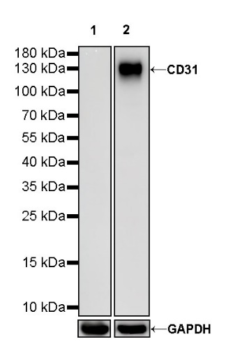
WB result of CD31 Mouse mAb
Primary antibody: CD31 Mouse mAb at 1/1000 dilution
Lane 1: HeLa whole cell lysate 20 µg
Lane 2: THP-1 whole cell lysate 20 µg
Negative control: HeLa whole cell lysate
Secondary antibody: Goat Anti-Mouse IgG, (H+L), HRP conjugated at 1/10000 dilution
Predicted MW: 82 kDa
Observed MW: 130 kDa
(This blot was developed with high sensitivity substrate)
Flow cytometric analysis of HeLa (Human cervix adenocarcinoma epithelial cell, left) / Jurkat (Human T cell leukemia T lymphocyte, right) cells labelling CD31 antibody at 1/200 dilution (1 μg)/ (Red) compared with a Mouse monoclonal IgG (Black) isotype control and an unlabelled control (cells without incubation with primary antibody and secondary antibody) (Blue). Goat Anti - Mouse IgG Alexa Fluor® 488 was used as the secondary antibody.
Negative control: HeLa
ICC shows positive staining in Jurkat cells. Anti-CD31 antibody was used at 1/100 dilution (Green) and incubated overnight at 4°C. Goat polyclonal Antibody to Mouse IgG - H&L (Alexa Fluor® 488) was used as secondary antibody at 1/1000 dilution. The cells were fixed with 4% PFA and permeabilized with 0.1% PBS-Triton X-100. Nuclei were counterstained with DAPI (Blue).
Negative control: ICC shows negative staining in HeLa cells. Anti-CD31 antibody was used at 1/100 dilution and incubated overnight at 4°C. Goat polyclonal Antibody to Mouse IgG - H&L (Alexa Fluor® 488) was used as secondary antibody at 1/1000 dilution. The cells were fixed with 4% PFA and permeabilized with 0.1% PBS-Triton X-100. Nuclei were counterstained with DAPI (Blue).
 您现在的位置: