
PBS, 40% Glycerol, 0.05% BSA, 0.03% Proclin 300
12 months from date of receipt / reconstitution, -20 °C as supplied
| 应用 | 稀释度 |
|---|---|
| Dot Blot | 1:1000 |
| WB | 1:1000 |
| IHC-P | 1:500 |
| IF | 1:500 |
| ChIP | 1:20~1:50 |
Acetylated histones, octameric proteins that organize chromatin into nucleosomes, the basic structural unit of the chromosomes and ultimately higher order structures, represent a type of epigenetic marker within chromatin. Acetylation removes the positive charge on the histones, thereby decreasing the interaction of the N termini of histones with the negatively charged phosphate groups of DNA. As a consequence, the condensed chromatin is transformed into a more relaxed structure that is associated with greater levels of gene transcription.
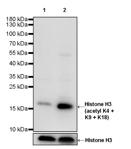
WB result of Histone H3 (acetyl K4 + K9 + K18) Rabbit pAb
Primary antibody: Histone H3 (acetyl K4 + K9 + K18) Rabbit pAb at 1/1000 dilution
Lane 1: HeLa whole cell lysate 20 µg
Lane 2: HeLa treated with Trichostatin A (100 ng/ml, 4 hour ) whole cell lysate 20 µg
Secondary antibody: Goat Anti-Rabbit IgG, (H+L), HRP conjugated at 1/10000 dilution
Predicted MW: 15 kDa
Observed MW: 17 kDa
Exposure time: 24 s
WB result of Histone H3 (acetyl K4 + K9 + K18) Rabbit pAb
Primary antibody: Histone H3 (acetyl K4 + K9 + K18) Rabbit pAb at 1/1000 dilution
Lane 1: NIH/3T3 whole cell lysate 20 µg
Lane 2: NIH/3T3 treated with Trichostatin A (100 ng/ml, 4 hour ) whole cell lysate 20 µg
Secondary antibody: Goat Anti-Rabbit IgG, (H+L), HRP conjugated at 1/10000 dilution
Predicted MW: 15 kDa
Observed MW: 17 kDa
Exposure time: 24 s
WB result of Histone H3 (acetyl K4 + K9 + K18) Rabbit pAb
Primary antibody: Histone H3 (acetyl K4 + K9 + K18) Rabbit pAb at 1/1000 dilution
Lane 1: C6 whole cell lysate 20 µg
Lane 2: C6 treated with Trichostatin A (100 ng/ml, 4 hour ) whole cell lysate 20 µg
Secondary antibody: Goat Anti-Rabbit IgG, (H+L), HRP conjugated at 1/10000 dilution
Predicted MW: 15 kDa
Observed MW: 17 kDa
Exposure time: 24 s
Dot blot result of Histone H3 (acetyl K4 + K9 + K18) Rabbit pAb
Lane 1: H3 (acetyl K18) peptide
Lane 2: H3 (acetyl K4) peptide
Lane 3: H3 (acetyl K9) peptide
Lane 4: H3 unmodified peptide
Primary antibody: Histone H3 (acetyl K4 + K9 + K18) Rabbit pAb at 1/1000 dilution
Secondary antibody: Goat Anti-Rabbit IgG, (H+L), HRP conjugated at 1/10000 dilution
Exposure time: 40 s
IHC shows positive staining in paraffin-embedded human testis. Anti-Histone H3 (acetyl K4 + K9 + K18) antibody was used at 1/500 dilution, followed by a HRP Polymer for Mouse & Rabbit IgG (ready to use). Counterstained with hematoxylin. Heat mediated antigen retrieval with Tris/EDTA buffer pH9.0 was performed before commencing with IHC staining protocol.
IHC shows positive staining in paraffin-embedded human breast cancer. Anti- Histone H3 (acetyl K4 + K9 + K18) antibody was used at 1/500 dilution, followed by a HRP Polymer for Mouse & Rabbit IgG (ready to use). Counterstained with hematoxylin. Heat mediated antigen retrieval with Tris/EDTA buffer pH9.0 was performed before commencing with IHC staining protocol.
IHC shows positive staining in paraffin-embedded human cervical squamous cell carcinoma. Anti- Histone H3 (acetyl K4 + K9 + K18) antibody was used at 1/500 dilution, followed by a HRP Polymer for Mouse & Rabbit IgG (ready to use). Counterstained with hematoxylin. Heat mediated antigen retrieval with Tris/EDTA buffer pH9.0 was performed before commencing with IHC staining protocol.
IHC shows positive staining in paraffin-embedded human thyroid cancer. Anti- Histone H3 (acetyl K4 + K9 + K18) antibody was used at 1/500 dilution, followed by a HRP Polymer for Mouse & Rabbit IgG (ready to use). Counterstained with hematoxylin. Heat mediated antigen retrieval with Tris/EDTA buffer pH9.0 was performed before commencing with IHC staining protocol.
IHC shows positive staining in paraffin-embedded mouse stomach. Anti- Histone H3 (acetyl K4 + K9 + K18) antibody was used at 1/500 dilution, followed by a HRP Polymer for Mouse & Rabbit IgG (ready to use). Counterstained with hematoxylin. Heat mediated antigen retrieval with Tris/EDTA buffer pH9.0 was performed before commencing with IHC staining protocol.
IHC shows positive staining in paraffin-embedded rat liver. Anti- Histone H3 (acetyl K4 + K9 + K18) antibody was used at 1/500 dilution, followed by a HRP Polymer for Mouse & Rabbit IgG (ready to use). Counterstained with hematoxylin. Heat mediated antigen retrieval with Tris/EDTA buffer pH9.0 was performed before commencing with IHC staining protocol.
IF shows positive staining in paraffin-embedded human stomach. Anti-Histone H3 (acetyl K4 + K9 + K18) antibody was used at 1/500 dilution (Green) and incubated overnight at 4°C. Goat polyclonal Antibody to Rabbit IgG - H&L (Alexa Fluor® 488) was used as secondary antibody at 1/1000 dilution. Counterstained with DAPI (Blue). Heat mediated antigen retrieval with EDTA buffer pH9.0 was performed before commencing with IF staining protocol.
Chromatin immunoprecipitation (ChIP) was performed on HeLa cells cross - linked with 1% formaldehyde for 10 min, then chromatin was fragmented by sonication. Parallel reactions used Histone H3 (acetyl K4 + K9 + K18) Rabbit polyclonal antibody and Rabbit mAb IgG Isotype Control (SDT-R173) at 1:50 for immunoprecipitation. Post - immunoprecipitation, both samples were washed, eluted, and cross - links reversed. Purified DNA was analyzed by qPCR.
qPCR (%input: immunoprecipitated DNA/input DNA) showed the enrichment of RPL30, GAPDH, MYOD1, AFM, SAT-α and SAT-2 in Histone H3 (acetyl K4 + K9 + K18) Rabbit polyclonal antibody - immunoprecipitated sample.
 您现在的位置: