
PBS, 40% Glycerol, 0.05% BSA, 0.03% Proclin 300
12 months from date of receipt / reconstitution, -20 °C as supplied
| 应用 | 稀释度 |
|---|---|
| Dot Blot | 1:1000 |
| WB | 1:1000 |
| ICC | 1:500 |
| ICFCM | 1:800 |
H3K79me2 is an epigenetic modification to the DNA packaging protein Histone H3. It is a mark that indicates the di-methylation at the 79th lysine residue of the histone H3 protein. H3K79me2 is detected in the transcribed regions of active genes.
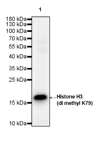
WB result of Histone H3 (di methyl K79) Rabbit pAb
Primary antibody: Histone H3 (di methyl K79) Rabbit pAb at 1/1000 dilution
Lane 1: HeLa whole cell lysate 20 µg
Secondary antibody: Goat Anti-Rabbit IgG, (H+L), HRP conjugated at 1/10000 dilution
Predicted MW: 17 kDa
Observed MW: 17 kDa
Exposure time: 3 s (This blot was developed with high sensitivity substrate)
WB result of Histone H3 (di methyl K79) Rabbit pAb
Primary antibody: Histone H3 (di methyl K79) Rabbit pAb at 1/1000 dilution
Lane 1: Neuro-2a whole cell lysate 20 µg
Secondary antibody: Goat Anti-Rabbit IgG, (H+L), HRP conjugated at 1/10000 dilution
Predicted MW: 17 kDa
Observed MW: 17 kDa
Exposure time: 3 s (This blot was developed with high sensitivity substrate)
Flow cytometric analysis of 4% PFA fixed 90% methanol permeabilized HeLa (Human cervix adenocarcinoma epithelial cell) labelling Histone H3 (di methyl K79) antibody at 1/800 dilution (0.1 μg)/ (Red) compared with a Rabbit monoclonal IgG (Black) isotype control and an unlabelled control (cells without incubation with primary antibody and secondary antibody) (Blue). Goat Anti - Rabbit IgG Alexa Fluor® 488 was used as the secondary antibody.
ICC shows positive staining in HeLa cells. Anti-Histone H3 (di methyl K79) antibody was used at 1/500 dilution (Green) and incubated overnight at 4°C. Goat polyclonal Antibody to Rabbit IgG - H&L (Alexa Fluor® 488) was used as secondary antibody at 1/1000 dilution. The cells were fixed with 100% ice-cold methanol and permeabilized with 0.1% PBS-Triton X-100. Nuclei were counterstained with DAPI (Blue). Counterstain with tubulin (Red).
Dot blot result of Histone H3 (di methyl K79) Rabbit pAb
Lane 1: H3 (di methyl K79) peptide
Lane 2: H3 (mono methyl K79) peptide
Lane 3: H3 (tri methyl K79) peptide
Lane 4: H3 unmodified peptide
Primary antibody: Histone H3 (di methyl K79) Rabbit pAb at 1/1000 dilution
Secondary antibody: Goat Anti-Rabbit IgG, (H+L), HRP conjugated at 1/10000 dilution
Exposure time: 90 s
 您现在的位置: