
12 months from date of receipt / reconstitution, -20 °C as supplied
| 应用 | 稀释度 |
|---|---|
| WB | 1:1000 |
| IP | 1:50 |
| IHC-P | 1:1000 |
| ICC | 1:500 |
Fascin, also known as 55 kDa actin-bundling protein, is an intracellular 55-58 kDa member of the fascin family of proteins. It has a restricted expression pattern, being found in oligodendrocytes, select endothelium, cerebellar stellate neurons, and blood. Fascin is found associated with actin in filopodia, and serves to coordinate and stabilize actin bundle formation, both in normal cells and tumor cells. Fascin expression is highly upregulated in many types of carcinomas including breast, colon, gastric, and oral squamous cell carcinomas. In these cancers, increased Fascin expression is linked to increased aggressiveness and high mortality. Thus, Fascin is a common biomarker for aggressive carcinomas, can be used as a histological marker of particular cancer subtypes, and may be a useful therapeutic target.
WB result of Fascin Rabbit mAb
Primary antibody: Fascin Rabbit mAb at 1/1000 dilution
Lane 1: HT-29 whole cell lysate 20 µg
Lane 2: HepG2 whole cell lysate 20 µg
Lane 3: HeLa whole cell lysate 20 µg
Negative control: HT-29 whole cell lysate
Secondary antibody: Goat Anti-Rabbit IgG, (H+L), HRP conjugated at 1/10000 dilution
Predicted MW: 55 kDa
Observed MW: 55 kDa
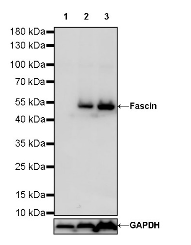
WB result of Fascin Rabbit mAb
Primary antibody: Fascin Rabbit mAb at 1/1000 dilution
Lane 1: NIH/3T3 whole cell lysate 20 µg
Lane 2: Neuro-2a whole cell lysate 20 µg
Secondary antibody: Goat Anti-Rabbit IgG, (H+L), HRP conjugated at 1/10000 dilution
Predicted MW: 55 kDa
Observed MW: 55 kDa
Fascin Rabbit mAb at 1/50 dilution (1 µg) immunoprecipitating Fascin in 0.4 mg HepG2 whole cell lysate.
Western blot was performed on the immunoprecipitate using Fascin Rabbit mAb at 1/1000 dilution.
Secondary antibody (HRP) for IP was used at 1/400 dilution.
Lane 1: HepG2 whole cell lysate 10 µg (Input)
Lane 2: Fascin Rabbit mAb IP in HepG2 whole cell lysate
Lane 3: Rabbit monoclonal IgG IP in HepG2 whole cell lysate
Predicted MW: 55 kDa
Observed MW: 55 kDa
IHC shows positive staining in paraffin-embedded human tonsil. Anti-Fascin antibody was used at 1/1000 dilution, followed by a HRP Polymer for Mouse & Rabbit IgG (ready to use). Counterstained with hematoxylin. Heat mediated antigen retrieval with Tris/EDTA buffer pH9.0 was performed before commencing with IHC staining protocol.
IHC shows positive staining in paraffin-embedded human kidney. Anti-Fascin antibody was used at 1/1000 dilution, followed by a HRP Polymer for Mouse & Rabbit IgG (ready to use). Counterstained with hematoxylin. Heat mediated antigen retrieval with Tris/EDTA buffer pH9.0 was performed before commencing with IHC staining protocol.
Negative control: IHC shows negative staining in paraffin-embedded human skeletal muscle. Anti-Fascin antibody was used at 1/1000 dilution, followed by a HRP Polymer for Mouse & Rabbit IgG (ready to use). Counterstained with hematoxylin. Heat mediated antigen retrieval with Tris/EDTA buffer pH9.0 was performed before commencing with IHC staining protocol.
IHC shows positive staining in paraffin-embedded human lung squamous cell carcinoma. Anti-Fascin antibody was used at 1/1000 dilution, followed by a HRP Polymer for Mouse & Rabbit IgG (ready to use). Counterstained with hematoxylin. Heat mediated antigen retrieval with Tris/EDTA buffer pH9.0 was performed before commencing with IHC staining protocol.
IHC shows positive staining in paraffin-embedded human malignant peripheral nerve sheath tumor (Tumor cells not positive). Anti-Fascin antibody was used at 1/1000 dilution, followed by a HRP Polymer for Mouse & Rabbit IgG (ready to use). Counterstained with hematoxylin. Heat mediated antigen retrieval with Tris/EDTA buffer pH9.0 was performed before commencing with IHC staining protocol.
IHC shows positive staining in paraffin-embedded human Hodgkin’s lymphoma. Anti-Fascin antibody was used at 1/1000 dilution, followed by a HRP Polymer for Mouse & Rabbit IgG (ready to use). Counterstained with hematoxylin. Heat mediated antigen retrieval with Tris/EDTA buffer pH9.0 was performed before commencing with IHC staining protocol.
IHC shows positive staining in paraffin-embedded mouse lung. Anti-Fascin antibody was used at 1/1000 dilution, followed by a HRP Polymer for Mouse & Rabbit IgG (ready to use). Counterstained with hematoxylin. Heat mediated antigen retrieval with Tris/EDTA buffer pH9.0 was performed before commencing with IHC staining protocol.
ICC shows positive staining in HeLa cells. Anti-Fascin antibody was used at 1/500 dilution (Green) and incubated overnight at 4°C. Goat polyclonal Antibody to Rabbit IgG - H&L (Alexa Fluor® 488) was used as secondary antibody at 1/1000 dilution. The cells were fixed with 100% ice-cold methanol and permeabilized with 0.1% PBS-Triton X-100. Nuclei were counterstained with DAPI (Blue). Counterstain with tubulin (Red).
Negative control: ICC shows negative staining in HT-29 cells. Anti-Fascin antibody was used at 1/500 dilution and incubated overnight at 4°C. Goat polyclonal Antibody to Rabbit IgG - H&L (Alexa Fluor® 488) was used as secondary antibody at 1/1000 dilution. The cells were fixed with 100% ice-cold methanol and permeabilized with 0.1% PBS-Triton X-100. Nuclei were counterstained with DAPI (Blue). Counterstain with tubulin (Red).
 您现在的位置: