
12 months from date of receipt / reconstitution, -20°C as supplied
| 应用 | 稀释度 |
|---|---|
| WB | 1:1000 |
| IP | 1:50 |
| IHC-P | 1:2000 |
PTP1B was first isolated from a human placental protein extract, but it is expressed in many tissues. It is localized to the cytoplasmic face of the endoplasmic reticulum. PTP1B can dephosphorylate the phosphotyrosine residues of the activated insulin receptor kinase. PTP1B is a negative regulator of the insulin signaling pathway and is considered a promising potential therapeutic target, in particular for treatment of type 2 diabetes. It has also been implicated in the development of breast cancer and has been explored as a potential therapeutic target in that avenue as well.
WB result of PTP1B Rabbit mAb
Primary antibody: PTP1B Rabbit mAb at 1/1000 dilution
Lane 1: HeLa whole cell lysate 20 µg
Lane 2: HCT 116 whole cell lysate 20 µg
Lane 3: HepG2 whole cell lysate 20 µg
Lane 4: A549 whole cell lysate 20 µg
Lane 5: MCF7 whole cell lysate 20 µg
Lane 6: SW480 whole cell lysate 20 µg
Secondary antibody: Goat Anti-Rabbit IgG, (H+L), HRP conjugated at 1/10000 dilution
Predicted MW: 50 kDa
Observed MW: 50 kDa
WB result of PTP1B Rabbit mAb
Primary antibody: PTP1B Rabbit mAb at 1/1000 dilution
Lane 1: RAW 264.7 whole cell lysate 20 µg
Secondary antibody: Goat Anti-Rabbit IgG, (H+L), HRP conjugated at 1/10000 dilution
Predicted MW: 50 kDa
Observed MW: 50 kDa
(This blot was developed with high sensitivity substrate)
PTP1B Rabbit mAb at 1/50 dilution (1 µg) immunoprecipitating PTP1B in 0.4 mg HCT116 whole cell lysate.
Western blot was performed on the immunoprecipitate using PTP1B Rabbit mAb at 1/1000 dilution.
Secondary antibody (HRP) for IP was used at 1/400 dilution.
Lane 1: HCT116 whole cell lysate 10 µg (Input)
Lane 2: PTP1B Rabbit mAb IP in HCT116 whole cell lysate
Lane 3: Rabbit monoclonal IgG IP in HCT116 whole cell lysate
Predicted MW: 50 kDa
Observed MW: 50 kDa
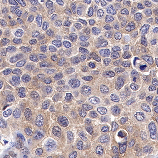
IHC shows positive staining in paraffin-embedded human bladder cancer. Anti-PTP1B antibody was used at 1/2000 dilution, followed by a HRP Polymer for Mouse & Rabbit IgG (ready to use). Counterstained with hematoxylin. Heat mediated antigen retrieval with Tris/EDTA buffer pH9.0 was performed before commencing with IHC staining protocol.
IHC shows positive staining in paraffin-embedded human lung cancer. Anti-PTP1B antibody was used at 1/2000 dilution, followed by a HRP Polymer for Mouse & Rabbit IgG (ready to use). Counterstained with hematoxylin. Heat mediated antigen retrieval with Tris/EDTA buffer pH9.0 was performed before commencing with IHC staining protocol.
IHC shows positive staining in paraffin-embedded human endometrial cancer. Anti-PTP1B antibody was used at 1/2000 dilution, followed by a HRP Polymer for Mouse & Rabbit IgG (ready to use). Counterstained with hematoxylin. Heat mediated antigen retrieval with Tris/EDTA buffer pH9.0 was performed before commencing with IHC staining protocol.
IHC shows positive staining in paraffin-embedded human pancreatic cancer. Anti-PTP1B antibody was used at 1/2000 dilution, followed by a HRP Polymer for Mouse & Rabbit IgG (ready to use). Counterstained with hematoxylin. Heat mediated antigen retrieval with Tris/EDTA buffer pH9.0 was performed before commencing with IHC staining protocol.
 您现在的位置: